
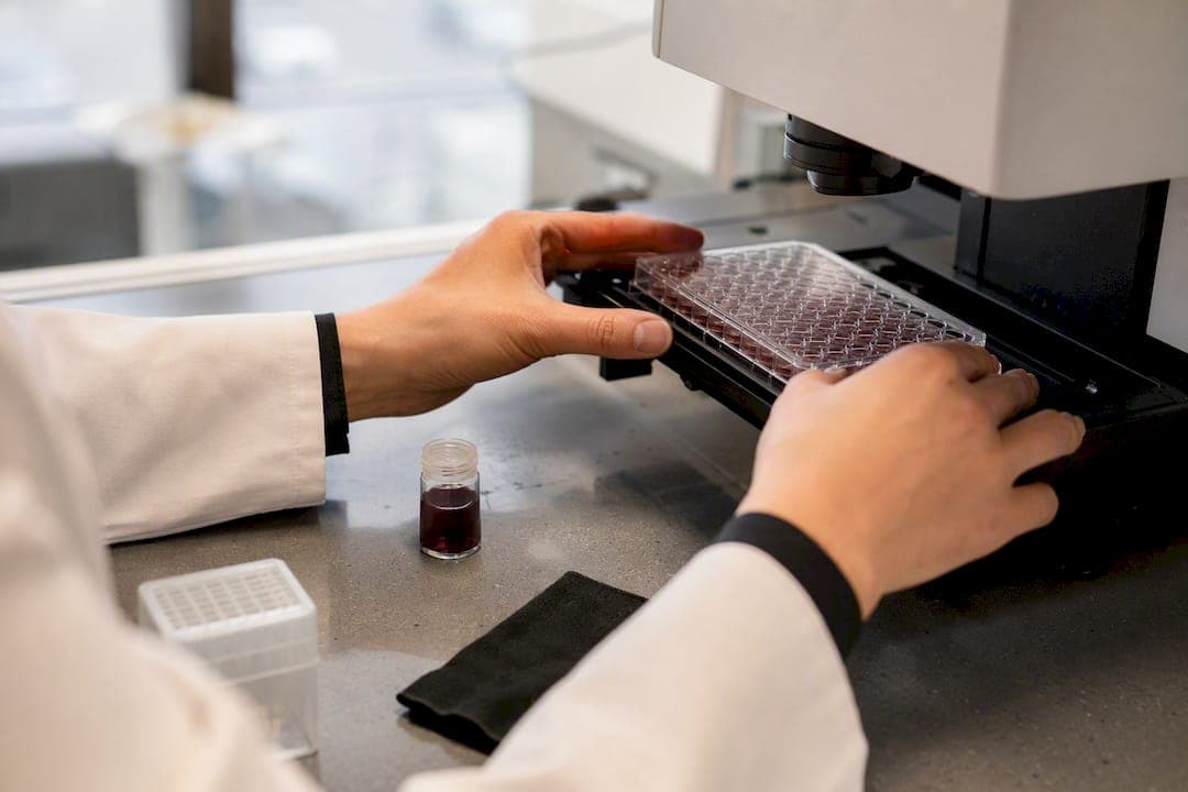
High content imaging (HCI) is an automated, image-based method that turns microscopy into multiparametric, decision-ready phenotypic data. It captures dozens of cellular features at once, size, shape, protein localization, texture, from thousands of samples, instead of measuring one endpoint per well. Use it when a single readout like viability or fluorescence intensity can't explain what's actually happening inside the cell.
- Best for: phenotypic profiling, mechanism-of-action studies, toxicity evaluation
- Core advantage: context-rich, multiparametric data instead of a single flat number
- Foundation: automated microscopy paired with quantitative image analysis
Key Takeaways
High content imaging works because it replaces single-endpoint measurements with multiparametric, single-cell phenotypic data that reveals mechanism, not just outcome.
| Point | Details |
|---|---|
| Definition matters | HCI extracts multiple phenotypic features per cell instead of one aggregate readout. |
| Workflow has three stages | Capture, analyze, and interpret each carry distinct technical decisions and failure points. |
| Instrument choice follows the question | Widefield suits throughput; confocal suits subcellular resolution and 3D structures. |
| QC needs multiple metrics | No single artifact check catches focus drift, saturation, and autofluorescence at once. |
| Hopeatrarelabs applies HCI to patient models | Multiparametric imaging profiles patient iPSC lines to rank drug and ASO candidates in parallel. |
Table of Contents
- The High Content Imaging Workflow: Capture, Analyze, Interpret
- Where High Content Imaging Fits: Applications and Assay Types
- Widefield vs Confocal: Choosing the Right Imaging Hardware
- Designing a Robust HCI Assay: Models, Controls, and QC
- How to Choose the Right HCI System or Partner
- A Real-World Example: HCI in Patient-Derived Disease Modeling
- What Comes Next for High Content Imaging
- Partnering With Hopeatrarelabs on High Content Imaging Projects
- Sources
The High Content Imaging Workflow: Capture, Analyze, Interpret
Every HCI project moves through three stages, and mistakes at any one of them ripple through the rest.
- Capture. You set channels, exposure times, z-stack depth, autofocus routines, and, for live samples, temperature and CO2 control. Get this wrong and no amount of downstream analysis fixes it.
- Analyze. Preprocessing corrects illumination and background noise, then segmentation identifies individual cells, nuclei, or organelles. Classical algorithms (watershed, thresholding) still work for clean samples, but deep-learning segmentation now handles the messy cases, touching cells, faint boundaries, dense colonies, far better.
- Interpret. Feature extraction produces a multiparametric profile per cell, which then feeds statistical comparison and visualization to separate real phenotypic shifts from noise.
Pro Tip: Budget analysis time separately from imaging time. Imaging can be the fastest part of the whole project once you factor in segmentation tuning, QC, and reanalysis cycles.
Raw image files pile up fast, and storage and compute routinely become the actual bottleneck, not the microscope. Plan your data architecture (raw retention policy, compressed intermediates, a reanalysis staging area) before the first plate goes on the stage, not after you've filled three hard drives.
Where High Content Imaging Fits: Applications and Assay Types
HCI earns its place in a workflow when the question is "how" or "why," not just "how much." A viability assay tells you cells died. HCI tells you whether they died from mitochondrial collapse, ER stress, or DNA damage, and that distinction changes which compound moves forward.
- Phenotypic screening and mechanism-of-action studies: compare morphological signatures against reference compounds with known targets to infer how an unknown hit works.
- Cell painting and morphological profiling: multiplexed fluorescent dyes stain multiple organelles simultaneously, generating a rich morphological fingerprint from a single well. Hopeatrarelabs walks through the staining and imaging steps in its protocol-first Cell Painting guide.
- Toxicology and organelle health: mitochondrial membrane potential, lysosomal accumulation, and nuclear fragmentation all show up as quantifiable image features well before a cell dies outright.
- 3D models, organoids, and small model organisms: spheroids and zebrafish larvae need z-stack imaging and segmentation tuned for depth and curvature, expanding HCI beyond flat monolayers.
Review evidence backs this breadth directly: HCI methods now cover toxicology, target validation, and novel endpoints across model organisms and 3D systems, which is a wider net than most researchers expect compared to what started as a 2D cell-counting tool. Related workflows for cell-based assay selection and drug screening build directly on these assay categories.
Widefield vs Confocal: Choosing the Right Imaging Hardware
Instrument choice comes down to a tradeoff between speed and optical cleanliness, and most labs get it wrong by defaulting to whichever system is already sitting in the core facility.
- Widefield imaging is fast and light-efficient, good for high-throughput screens where you need thousands of wells imaged quickly and z-resolution isn't critical.
- Confocal imaging rejects out-of-focus light for cleaner optical sections, essential for 3D structures, thick spheroids, or any assay where subcellular localization is the actual readout.
- Detectors matter more than most people budget for: sCMOS sensors dominate modern HCI for their speed and dynamic range, CCDs still show up in legacy systems, and PMTs remain standard in confocal laser-scanning setups.
- Illumination: LED sources are cheaper and last longer; lasers deliver higher intensity for confocal and are close to mandatory for fast live-cell work.
- Automation and environmental control: plate handlers and incubation chambers become non-negotiable once you're running live-cell kinetics or scaling past a few dozen plates per week.
Pro Tip: If you're torn between widefield and confocal, ask what you're measuring, not what looks nicer. A texture or intensity readout rarely needs confocal resolution; subcellular colocalization almost always does.
Designing a Robust HCI Assay: Models, Controls, and QC
Assay quality is decided before the microscope ever turns on. The Assay Guidance Manual makes a point that's easy to underweight: biological model, reagents, and analysis strategy matter as much as the imaging hardware itself.
- Pick the right biological model. 2D immortalized lines are cheap and fast but often miss disease-relevant biology. Primary cells and iPSC-derived models cost more time and money but capture patient-specific phenotypes that 2D lines simply can't. 3D models add physiological relevance at the cost of imaging complexity.
- Plan your labeling strategy before you plate anything. Spectral overlap and autofluorescence are the two most common reasons a promising assay produces garbage data. Map your channels against your dyes' actual emission spectra, not just their marketing names.
- Build in controls from day one. Reference compounds with known phenotypic signatures let you confirm the assay is working before you trust a single hit. Counter-screens catch compound interference, and autofluorescence and quenching artifacts are common sources of false positives and negatives that orthogonal follow-up assays are designed to catch.
- Watch for the usual failure modes. Focus drift, saturation, debris, and edge effects account for most technical noise in HCI datasets. No single QC metric catches everything; experienced practitioners combine several artifact-specific metrics or supervised classifiers rather than relying on one threshold.
- Size your compute and storage before you scale. A pilot run on 96 wells behaves nothing like a 384-well screen once raw image volume multiplies. Reanalysis staging areas save real time later.
Hopeatrarelabs' guides on validating cellular disease models and prioritizing drug candidates from patient iPSCs go deeper into steps 1 and 3 for anyone building assays around patient-derived models specifically.
How to Choose the Right HCI System or Partner
Start with your decision point, not your budget. Early discovery work tolerates lower resolution in exchange for higher throughput; mechanism-of-action and toxicity follow-up usually need better optics and slower, more careful imaging.
- Sensitivity and resolution should match your smallest phenotypic difference, not your largest.
- Throughput determines whether you need full plate automation or can manage with manual loading.
- Analysis software and AI features vary widely; some platforms now bundle AI-powered segmentation and phenotypic classification directly into the acquisition software, cutting manual tuning time substantially, though results are only as good as the training data behind them.
- Vendor support and expandability matter more once you're two years in and need new modules, not on day one.
- Outsourcing to a core facility or contract lab makes sense when the scale or specialized expertise (3D imaging, rare disease models, custom analysis pipelines) exceeds what an in-house setup can justify. Compare the throughput tradeoffs of HCS vs HCI before committing to either path.
A Real-World Example: HCI in Patient-Derived Disease Modeling
Hopeatrarelabs runs HCI at the center of its rare disease work, not as an add-on. The workflow starts with a patient sample, converts it into a disease-specific induced pluripotent stem cell (iPSC) line, and then applies image-based assays to characterize the disease phenotype at the cellular level.
- Multiparametric imaging profiles morphology, organelle health, and protein localization across the patient's cells versus CRISPR-corrected isogenic controls.
- Those phenotypic signatures guide hit selection when testing thousands of approved drugs and custom ASOs in parallel against the same model.
- Top candidates get ranked by phenotypic rescue strength, then move into orthogonal validation before any translational discussion begins.
- Protocol-level detail on the imaging assays behind this work, including autophagy assays in iPSC models, is published for researchers building similar pipelines.
What Comes Next for High Content Imaging
AI-driven segmentation is closing the gap between manual expert annotation and automated phenotyping, though it still lives or dies on training data quality. Expect tighter integration with multiomics and continued growth in 3D imaging fidelity over the next few years, trends we track in more depth in our biopharma innovation trends piece.
If you're starting out, run a small pilot before committing to a full screen, and nail down your QC metrics before you generate data you'll need to throw away. Hopeatrarelabs publishes protocol-level resources for labs building these pipelines from scratch.
— John
Partnering With Hopeatrarelabs on High Content Imaging Projects
Hopeatrarelabs is the option for rare disease programs that need patient-specific answers, not just a general screening platform. Where a standard core facility runs your plates and hands back raw data, Hopeatrarelabs builds the entire pipeline around one patient's biology: a CRISPR-edited isogenic iPSC model, imaging-based phenotypic assays tuned to that specific disease, and parallel screening across thousands of approved drugs and custom ASOs in a single coordinated program.
That matters most when a disease is too rare for an off-the-shelf assay to exist yet, or when a family or foundation needs a translational report they can actually bring to a physician. Running this in-house demands iPSC expertise, imaging infrastructure, and analysis pipelines most labs don't have sitting idle. If you're a patient, family, foundation, or biopharma partner facing an undiagnosed or ultra-rare genetic disease, visit the Hopeatrarelabs landing page to start a conversation about a personalized screening program.
Sources
- High-Content Imaging & Analysis | HCI Systems | Molecular Devices
- Assay Development Guidelines for Image-Based High Content Screening, High Content Analysis and High Content Imaging - Assay Guidance Manual - NCBI Bookshelf
- Applications of High Content Screening in Life Science Research - PMC