

Validate a cellular disease model by predefining its Context of Use (COU), setting measurable acceptability criteria, and demonstrating concordant evidence across genomic, phenotypic, and functional axes with quantified uncertainty. That sequence is not optional. The Ten Rules for credible practice, ASME V&V 40, and FDA credibility frameworks all converge on the same starting point: rigor scales with COU, and post-hoc rationalization is the single most common failure mode in cellular model validation.
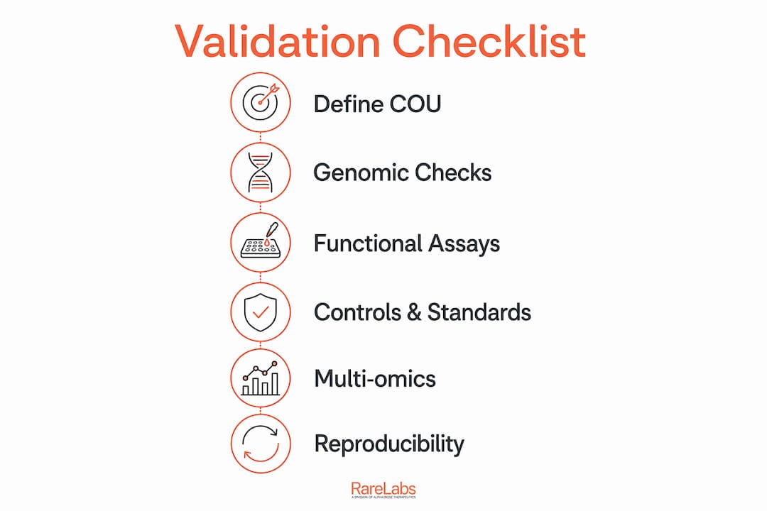
Minimum validation checklist:
- COU statement: Written, pre-specified, and tied to a decision (research use, target prioritization, translational assay, or regulatory support)
- Identity and authenticity: STR profiling or WGS-derived SNV concordance >99% to donor; karyotype for structural stability
- Genomic and transcriptomic benchmarks: Karyotype, WGS/WES or targeted panel, bulk RNA-seq for average expression, scRNA-seq for rare or off-target cell types
- Phenotypic endpoints: Protein marker expression (IHC/ICC, flow cytometry), morphology by imaging, electrophysiology where COU requires it
- Functional readouts: Drug-response curves (EC50/IC50), pathway activity assays, rescue experiments (genetic or pharmacologic)
- Controls: Isogenic controls, healthy-donor controls, positive/negative perturbation controls, vehicle controls
- Reproducibility: Pre-specified biological replicate counts, power analysis documented before data collection, effect sizes reported
- Uncertainty quantification: Bootstrap or Bayesian credible intervals on key predictions; sensitivity analysis identifying dominant input drivers
- Documentation: Versioned SOPs, reagent lot numbers, instrument settings, raw data accession IDs, and a completed reporting checklist
A model that captures disease-relevant features — histology, gene regulation, protein expression, pharmacological responses — and compares outputs to independent data without post-hoc parameter tuning meets the baseline bar. Everything below expands each checkpoint into executable steps.
Table of Contents
- 1. Define context of use before you design a single assay
- 2. How to run genomic and molecular identity checks
- 3. Phenotypic and functional assays: orthogonal evidence every model should provide
- 4. Controls, reference standards, and benchmarking strategy
- 5. Multi-omics and spatial methods to validate architecture and cell composition
- 6. Robustness, reproducibility, uncertainty quantification, and sensitivity analysis
- 7. Generating and maintaining stem cell-derived disease models: practical QC steps
- 8. Reporting, metadata standards, and data sharing for reproducibility
- 9. Practical validation checklist, timeline, and resource considerations
- 10. How Hopeatrarelabs validates patient-specific cellular disease models
- Key takeaways
- The part of validation most researchers underestimate
- Hopeatrarelabs supports validation from first cell to final report
- Selected authoritative resources and standards to read next
1. Define context of use before you design a single assay
The COU is the primary trust anchor for any validation package. It answers: What decision will this model inform, and under what conditions? A model used to generate exploratory mechanistic hypotheses needs far less evidence than one supporting a preclinical go/no-go decision or a regulatory submission.
Write the COU as a single declarative sentence before any data collection begins. For example: "This iPSC-derived cardiomyocyte model will be used to rank-order cardiotoxicity risk of ten candidate compounds in a drug-discovery screen." That sentence immediately tells you which endpoints matter (electrophysiology, viability), what comparators are needed (positive control compound, healthy-donor line), and what statistical threshold constitutes a pass.
Pro Tip: Register your COU and acceptability criteria in a lab notebook or electronic system before the first experiment. Reviewers and collaborators can then verify that thresholds were not moved after results were seen — a practice aligned with the Ten Rules conformance rubric and essential for any regulatory-adjacent use.
Concrete COU examples and the evidence each demands:
| COU | Key evidence required |
|---|---|
| Drug-response screening | EC50/IC50 concordance, positive control response, replicate CV |
| Mechanistic hypothesis test | Pathway assay, rescue experiment, orthogonal readout |
| N-of-1 treatment prioritization | Patient-matched isogenic control, functional + genomic concordance |
| Regulatory translational support | Full V&V package per ASME V&V 40 / FDA credibility framework |
2. How to run genomic and molecular identity checks
Start with identity before anything else. A model that drifts genetically or carries contamination will produce unreproducible data regardless of how well the downstream assays are designed.
Core identity and contamination checks:
- STR profiling or WGS SNV concordance: Confirm >99% match to the donor source. Run at establishment and after any major passage event or cryorecovery.
- Karyotype: G-banding or array CGH at minimum; repeat after extended culture to catch structural instability introduced by reprogramming or differentiation stress.
- Mycoplasma testing: PCR-based assay every 4–8 weeks and after any new reagent lot introduction. Mycoplasma silently alters gene expression and drug response.
- Cross-contamination screen: STR or SNP panel against a panel of common cell lines (ATCC authentication service or equivalent) at establishment.
For transcriptomic benchmarking, bulk RNA-seq gives you average expression across the population and is the workhorse for comparing your model to reference tissue datasets. Single-cell RNA-seq adds the resolution to detect rare off-target cell types that bulk methods miss entirely — a contaminating 5% fibroblast population is invisible in bulk but obvious in a UMAP. Advanced validation pairs bulk RNA-seq with scRNA-seq specifically because each answers a different question.
WGS is preferred over targeted panels when the disease mechanism involves structural variants or when the model is intended for translational or regulatory use. For exploratory research, a well-designed targeted panel with documented variant calling pipeline, genome build (GRCh38), and software version is usually sufficient. Document all of this in the methods section and data accession record.

3. Phenotypic and functional assays: orthogonal evidence every model should provide
No single assay should carry the validation decision alone. The logic is straightforward: any one readout can be artifactual, and two independent assays converging on the same biological claim is qualitatively stronger evidence than one assay run at higher throughput.
Design your functional tests to map directly to the COU. A cardiomyocyte model for arrhythmia research needs multi-electrode array (MEA) recordings and patch-clamp data, not just protein markers. A neuronal model for synaptic disease needs electrophysiology and calcium imaging, not just morphology. Morphology alone is necessary but never sufficient.
Pro Tip: Validate functional endpoints with blinded scoring and automated image analysis pipelines (e.g., CellProfiler for morphology, automated MEA analysis software) before unblinding treatment conditions. Observer bias in manual scoring is a reproducibility killer that rarely appears in methods sections but frequently explains why results do not replicate across labs.
Rescue experiments are underused and undervalued. If the disease phenotype disappears when you correct the causal variant (CRISPR correction) or add back the missing protein, that is the strongest functional evidence available. Pharmacologic rescue with a known-mechanism compound is the next best option. Both should be pre-specified in the COU document as required evidence, not optional add-ons.
For drug-response validation, report full dose-response curves with EC50/IC50 values, Hill coefficients, and confidence intervals. A single-concentration readout is not a dose-response curve. Concordance between the model's pharmacological response and published clinical or in vivo data is the translational bridge that clinical anchoring of assays requires.
4. Controls, reference standards, and benchmarking strategy
Controls are not a formality. They are the interpretive framework that makes your data meaningful.
Essential controls for every validation experiment:
- Isogenic controls: Same genetic background as the disease line, with the causal variant corrected. This is the gold standard comparator for iPSC-derived models.
- Healthy-donor controls: At least two independent donors to separate disease signal from donor-specific variation.
- Positive perturbation controls: A compound or genetic manipulation with a known, reproducible effect on the endpoint being measured.
- Negative/vehicle controls: DMSO at the same concentration as the highest drug treatment, or a non-targeting siRNA/sgRNA.
- Technical blanks: No-cell wells or empty vector controls to quantify background signal.
For benchmarking against public datasets, document the reference dataset version, the tissue atlas used (Human Cell Atlas, GTEx, ENCODE), and the date of access. Benchmark metrics worth reporting include sensitivity, specificity, and distributional concordance measures such as KL divergence or Wasserstein distance when comparing transcriptomic profiles.
One trap to avoid: circular benchmarking, where model parameters are tuned to match a single reference dataset and then that same dataset is used to claim validation. Independent validation requires a dataset the model has never seen during development.
5. Multi-omics and spatial methods to validate architecture and cell composition
For 2D monolayer models, bulk RNA-seq plus scRNA-seq usually covers the validation need. For 3D organoids, co-cultures, and organ-on-chip systems, spatial transcriptomics or spatial proteomics is often required to demonstrate that the architecture relevant to the COU is actually preserved.
Why spatial methods change the interpretation: A cerebral organoid may express the correct neuronal markers in bulk RNA-seq and still have its cell types distributed in a biologically incorrect spatial pattern. Spatial transcriptomics (10x Visium, Slide-seq, or equivalent) reveals whether excitatory neurons, inhibitory interneurons, and glia are organized in a tissue-relevant arrangement — or randomly intermixed. That distinction matters for any COU involving cell-cell signaling, synaptic connectivity, or drug penetration gradients.
Multi-omics approaches — bulk RNA-seq for average expression, scRNA-seq for rare populations, ATAC-seq for chromatin accessibility, and spatial transcriptomics for architecture — are not redundant. Each layer answers a question the others cannot. Proteomics adds post-translational context that transcript data misses entirely.
Practical sampling considerations: for scRNA-seq, aim for a minimum of 5,000 cells per sample to reliably detect populations present at 1–2% frequency. Batch design matters enormously; process disease and control samples in the same sequencing run wherever possible, and document batch assignments in the metadata. ATAC-seq requires fresh or flash-frozen material and degrades rapidly with poor handling.
6. Robustness, reproducibility, uncertainty quantification, and sensitivity analysis
Reproducibility is not a post-publication concern. It needs to be designed in from the start.
Statistical and practical requirements:
- Replicate structure: Distinguish technical replicates (same sample, repeated measurement) from biological replicates (independent differentiations or donors). Power analyses should be based on biological replicates, not technical ones.
- Pre-specified endpoints: Lock down primary and secondary endpoints in the COU document before data collection. Moving endpoints after seeing data is post-hoc rationalization.
- Effect size reporting: Report Cohen's d or equivalent alongside p-values. A statistically significant result with a trivial effect size is not biologically meaningful.
- Uncertainty quantification: Use bootstrap confidence intervals, Monte Carlo sampling, or Bayesian credible intervals for key predictions. For computational models, closed-loop validation pairs computational uncertainty estimates with experimental corroboration.
- Sensitivity analysis: Identify which input variables drive the most output variance. This guides where to invest experimental controls and where model conclusions are fragile.
Pro Tip: Automating routine liquid handling and cell seeding early in your workflow reduces human-induced variance, which is often the dominant source of inter-lab irreproducibility. A Hamilton or Tecan liquid handler for media changes and compound addition pays for itself in reproducibility within a single multi-site study.
Validation is an iterative cycle. When a model fails to reproduce the expected biology, return to the conceptual design rather than adjusting parameters to force a fit. Overfitting to a single dataset produces a model that looks validated on paper and fails on the next independent dataset.
7. Generating and maintaining stem cell-derived disease models: practical QC steps
iPSC-based models introduce QC requirements that primary cell models do not have. The reprogramming step itself can introduce genomic instability, and differentiation efficiency varies between batches in ways that are not always visible without staged marker checks.
The most common mistake in iPSC model generation is treating a successful differentiation as proof of a validated model. Differentiation efficiency and marker expression confirm that the protocol worked. They do not confirm that the resulting cells recapitulate the disease biology. Those are two separate questions requiring separate evidence.
Starting material and reprogramming QC:
- Confirm donor consent and sample provenance documentation before any work begins.
- Run karyotype on iPSC lines before differentiation, not after. Aneuploid lines waste months of downstream work.
- Sequence the causal variant to confirm it is present (disease lines) or corrected (isogenic controls) at the iPSC stage.
Differentiation QC and maintenance:
- Use staged marker expression checkpoints at each differentiation step, with pre-defined pass/fail criteria (e.g., >80% NKX2.5+ at cardiac progenitor stage).
- Set passage limits and document them in the SOP. Most iPSC lines show increased genomic instability beyond passage 50.
- Test for mycoplasma at every cryorecovery. Log lot numbers for all media components and growth factors, since batch variation in recombinant proteins is a real and underappreciated source of differentiation variability.
- Document failed differentiation routes explicitly. A failed attempt that is not recorded will be repeated by the next researcher on the project.
The ISSCR standards for stem cell-based model systems provide the reference framework for iPSC model governance, including guidelines on authentication, banking, and quality release criteria.
8. Reporting, metadata standards, and data sharing for reproducibility
A validation package that cannot be reproduced from the methods section is not a validation package. It is a result.
Minimum reporting checklist:
- COU statement (verbatim from the pre-specified document)
- Protocol version and SOP identifier for each assay
- Reagent lot numbers, catalog numbers, and supplier for all critical materials
- Instrument model, firmware/software version, and calibration date
- Raw data accession identifiers (GEO for sequencing, Zenodo or Figshare for imaging data)
- Genome build and variant calling pipeline version for any sequencing data
- Statistical analysis code (GitHub repository or container image with version tag)
- Batch assignment table for all multi-batch experiments
For computational model components, SBML and CellML are the standard encoding formats. MIRIAM-compliant annotation links model components to database identifiers (UniProt, ChEBI, GO) and makes models reusable by others. The ASME V&V 40 and FDA credibility framework provide the formal structure for verification and validation documentation in regulatory-adjacent contexts.
Sequencing data minimum metadata: organism, tissue/cell type, passage number, treatment conditions, library preparation kit and version, sequencer model, and read depth. FASTQ files go to SRA or ENA with a BioProject accession. Processed matrices go to GEO. Neither replaces the other.
9. Practical validation checklist, timeline, and resource considerations
Phased validation timeline:
- Planning (weeks 1–2): Write COU, define endpoints, pre-specify acceptability criteria, assign roles, identify reference datasets and controls.
- Pilot (weeks 3–6): Run endpoint assays at small scale to estimate variance, refine protocols, and conduct power analysis for the powered validation run.
- Powered validation (weeks 7–16): Execute pre-specified experiments with full replicate structure, blinded where feasible.
- Cross-platform replication (weeks 17–24): Replicate key findings in an independent lab or on an independent instrument platform.
- Documentation and reporting (weeks 25–28): Compile reporting checklist, deposit data, finalize SOPs.
| Task | Estimated duration | Resource intensity | Responsible role |
|---|---|---|---|
| COU definition and criteria | 1–2 weeks | Low | PI, statistician |
| Karyotype + STR authentication | 1–2 weeks | Low–medium | Assay scientist |
| Bulk RNA-seq + analysis | 3–5 weeks | Medium | Assay scientist, bioinformatician |
| scRNA-seq + analysis | 4–8 weeks | High | Assay scientist, bioinformatician |
| Phenotypic assays (IHC, flow) | 2–4 weeks | Medium | Assay scientist |
| Electrophysiology (MEA/patch) | 3–6 weeks | High | Specialist |
| Drug-response screening | 3–5 weeks | Medium–high | Assay scientist |
| Cross-platform replication | 6–10 weeks | High | External partner or second lab |
| Documentation and data deposit | 2–4 weeks | Low–medium | QA, bioinformatician |
Involve a statistician at the planning stage, not after data collection. Bioinformaticians need to be part of the sequencing study design, not just the analysis. External reviewers for independent replication are worth the cost for any model intended for translational or regulatory use.
10. How Hopeatrarelabs validates patient-specific cellular disease models
Hopeatrarelabs builds patient-specific disease models for ultra-rare and undiagnosed genetic diseases, using iPSC reprogramming and CRISPR gene editing as the core platform. The validation workflow maps directly onto the checklist above, with explicit pass/fail gates at each stage.
The Hopeatrarelabs pipeline treats each checkpoint as a hard gate, not a soft recommendation. A line that fails karyotype does not proceed to differentiation. A differentiation batch that misses staged marker thresholds is rejected and the protocol is reviewed before the next attempt. Documentation is recorded in versioned SOPs with data accession identifiers at each stage.
The workflow runs as follows: patient sample acquisition and consent verification → iPSC reprogramming with genomic integrity check (karyotype, causal variant confirmation) → staged differentiation with marker-based release criteria → functional screening using parallel drug screens across FDA-approved compounds, custom ASOs, and gene therapy options → orthogonal functional assays to build convergent evidence → prioritized treatment recommendations with documented uncertainty bounds.
Parallel drug screens are a deliberate design choice. Testing thousands of compounds simultaneously against the patient's own cells, rather than sequentially, compresses the timeline and generates dose-response data across a pharmacological space that sequential screening cannot cover. Orthogonal functional assays — at minimum two independent readouts per biological claim — prevent any single assay artifact from driving a treatment recommendation. For researchers interested in cellular models in drug discovery or patient-derived cell applications, the Hopeatrarelabs knowledge base provides additional protocol context.
Key takeaways
Cellular model validation succeeds when COU is defined first, acceptability criteria are pre-specified, and at least two orthogonal readouts converge on the same biological claim with quantified uncertainty.
| Point | Details |
|---|---|
| COU drives everything | Write the Context of Use before any experiment; it determines which assays, controls, and thresholds are required. |
| Orthogonal readouts are required | No single assay should carry the validation decision; require at least two independent endpoints that converge. |
| Pre-specify thresholds | Lock acceptability criteria before data collection to prevent post-hoc rationalization. |
| Quantify uncertainty | Report bootstrap or Bayesian credible intervals on key predictions and run sensitivity analysis on dominant input drivers. |
| Hopeatrarelabs exemplar | Hopeatrarelabs implements hard pass/fail gates at each validation checkpoint, using parallel drug screens and orthogonal functional assays for translational decision-making. |
The part of validation most researchers underestimate
The field has gotten good at describing validation. It has not gotten good at doing it before the data looks interesting.
The most common failure pattern is not a missing assay. It is a researcher who runs the assays, sees a result that looks compelling, and then writes the acceptability criteria to match what they observed. That is not validation. It is confirmation bias with a methods section. The pre-specification requirement exists precisely because humans are not good at evaluating evidence they generated themselves.
The second underestimated problem is biological replicate count. Most labs run three biological replicates because that is what fits on a plate. Three replicates is almost never enough to detect a biologically meaningful effect size with adequate power, especially in iPSC models where inter-differentiation variance is high. Run a power analysis. If the answer is uncomfortable, that is the point.
Simpler, well-validated systems are often more translatable than complex ones. More complexity does not guarantee better prediction — a 2D model with rigorous controls and pre-specified endpoints frequently outperforms a 3D organoid with neither. Build complexity only when the COU genuinely requires it, and validate the simpler version first.
Finally: document your failures. A failed differentiation route that is not recorded will be repeated. A negative drug-response result that is not deposited will be rediscovered at cost by someone else. The GPAT framework for model transportability is a useful reminder that even well-characterized in vitro models have limited transportability to in vivo phenotypes — and that knowing the limits of your model is as valuable as knowing its strengths.
Hopeatrarelabs supports validation from first cell to final report
For research teams working on ultra-rare or undiagnosed genetic diseases, the gap between knowing the validation checklist and having the infrastructure to execute it is real. Hopeatrarelabs offers iPSC-based model development, genomic and functional QC pipelines, and parallel drug screens built around the COU-driven framework described in this article.

Every engagement starts with a defined COU and pre-specified acceptability criteria, then moves through genomic authentication, staged differentiation QC, and orthogonal functional screening. The output is a documented validation package with versioned SOPs and data accession records, not just a results summary. For teams needing gene therapy screening or parallel compound screening against a patient-specific model, Hopeatrarelabs operates as a scientific partner, not a contract lab. Contact the team at hopeatrarelabs.com to discuss a pilot validation engagement or collaboration.
Selected authoritative resources and standards to read next
These are the primary standards and reviews cited in this article. Cite them directly when documenting validation in manuscripts or regulatory submissions.
- Ten Rules for credible practice of modeling and simulation (Nature Reviews Bioengineering): The foundational COU-first framework; defines how rigor scales with intended use and provides the vocabulary for validation documentation.
- Conformance rubric for the Ten Rules (PLOS ONE): A practical scoring tool for assessing how well a validation package meets each of the Ten Rules; useful for self-assessment before submission.
- ASME V&V 40 / FDA credibility framework (Journal of Translational Medicine): Adapts engineering V&V standards to computational systems biology; covers code verification, calculation verification, and COU-based credibility goals.
- Screening out irrelevant cell-based models (Nature Reviews Drug Discovery): Argues for clinical anchoring of assay design and multidisciplinary collaboration; essential reading for translational validation.
- AI-driven virtual cell models: closed-loop validation (npj Digital Medicine): Covers uncertainty quantification and experimental corroboration in computational-experimental validation loops.
- GPAT framework for model transportability (PMC): Gene perturbation-based method for assessing whether in vitro cellular phenotypes transport to in vivo human outcomes; useful for translational relevance assessment.
- ISSCR standards for stem cell-based model systems: Reference governance framework for iPSC model authentication, banking, and quality release.
- Peptides in rare disease research (Peptilab): Partner resource covering peptide-based assay tools relevant to researchers using peptide probes or therapeutic peptides in disease model validation.