

Genetic anatomy refers to the physical and regulatory organization of a gene: the ordered sequence of elements that determine when, where, and how that gene produces its product. The term isn't standard clinical vocabulary the way "genomics" or "molecular genetics" are, but it captures something real and useful: genes have architecture, and that architecture is the difference between a gene that works and one that causes disease.
The core verdict: gene function depends on far more than the protein-coding sequence. The surrounding regulatory landscape, the splice signals, the epigenetic marks, and the three-dimensional folding of chromatin all shape what a gene actually does in a living cell.
Here's what this guide covers:
- The key terms you need: gene, genome, chromosome, allele, exon, intron, UTR
- The ordered parts of a typical protein-coding gene and their roles
- Coding versus non-coding DNA and why the distinction matters
- How genes are regulated by promoters, enhancers, and epigenetic marks
- How genes are read: transcription and translation, step by step
- Alternative splicing and why one gene can make many proteins
- How variants in gene anatomy cause disease
- The methods scientists use to study gene structure and expression
- Why spatial gene expression maps matter for patients and clinicians
Key Takeaways
Genetic anatomy, the ordered structural and regulatory organization of a gene, determines not just what a gene encodes but when, where, and how much of its product is made, and variants anywhere in that architecture can drive disease.
| Point | Details |
|---|---|
| Genes have ordered architecture | Each gene contains a promoter, UTRs, exons, introns, splice sites, and a poly-A signal, each with a distinct role. |
| Less than 2% codes for protein | The vast majority of the human genome is non-coding and includes regulatory elements, ncRNA genes, and structural sequences. |
| Regulation extends beyond sequence | Enhancers, silencers, DNA methylation, and histone modifications control gene activity independently of the coding sequence. |
| One gene, many proteins | Alternative splicing allows a single gene to produce multiple protein isoforms; splice-site variants are a common and underdiagnosed disease mechanism. |
| Spatial expression shapes interpretation | Knowing where a gene is expressed helps connect variants to affected tissues and guides patient-specific disease modeling. |
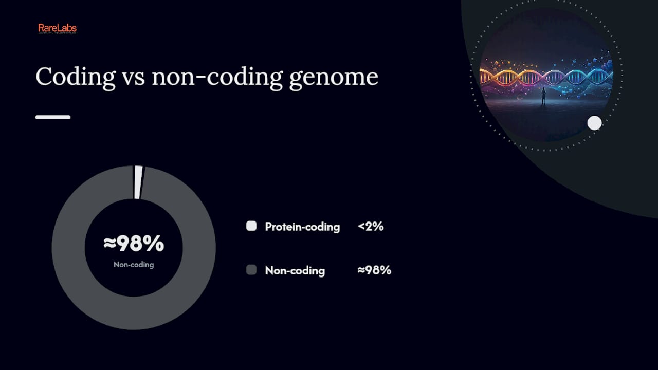
Table of Contents
- What do the key genetics terms actually mean?
- What are the structural parts of a genetic anatomy?
- What counts as coding DNA versus non-coding DNA?
- How do promoters, enhancers, and epigenetics regulate genes?
- How does a gene get read? Transcription and translation
- Why can one gene produce many different proteins?
- How do changes in gene anatomy lead to disease?
- What methods do scientists use to study gene anatomy?
- Why spatial gene expression maps matter for patients and clinicians
- Why understanding gene anatomy matters for patients and families
- An editorial perspective on genetic anatomy and rare disease
- Sources
What do the key genetics terms actually mean?
Before going further, a short vocabulary map. These terms appear throughout any discussion of gene structure, and the definitions below are written for clarity, not comprehensiveness.
- Gene: A segment of DNA that encodes a functional product, usually a protein or a non-coding RNA. NCBI's genetics primer defines it as the basic physical and functional unit of heredity.
- Genome: The complete set of DNA in an organism, including all genes and non-coding sequences. The human genome contains roughly 3 billion base pairs.
- Chromosome: A tightly packaged structure of DNA and protein. Humans have 46 chromosomes, arranged in 23 pairs.
- Allele: One version of a gene. You carry two alleles of most genes, one from each parent. Eye color is the classic example: different alleles at the same chromosomal location produce different pigmentation outcomes.
- Exon: The portion of a gene that ends up in the final, processed messenger RNA (mRNA) and contributes to the protein or functional RNA product.
- Intron: A sequence within a gene that is transcribed but then removed (spliced out) before the mRNA is used. Introns are often far longer than exons.
- 5' UTR (5' untranslated region): The stretch of mRNA upstream of the start codon. It helps position the ribosome and can regulate translation efficiency.
- 3' UTR (3' untranslated region): The stretch of mRNA downstream of the stop codon. It contains signals for mRNA stability, localization, and polyadenylation.
- Promoter: A DNA sequence just upstream (5') of a gene where RNA polymerase and transcription factors bind to initiate transcription.
- Enhancer: A regulatory DNA element that can boost transcription of a target gene, sometimes from tens of thousands of base pairs away.
- Splice site: The short consensus sequences at the boundaries of introns (GT at the 5' end, AG at the 3' end) that guide the splicing machinery.
- Transcription factor: A protein that binds specific DNA sequences to activate or repress gene transcription.
Allele and trait, connected: The HERC2/OCA2 region on chromosome 15 contains alleles that control the amount of melanin produced in the iris. A single nucleotide change in an enhancer within HERC2 reduces OCA2 expression, shifting eye color from brown toward blue. One regulatory variant, one measurable trait.
What are the structural parts of a genetic anatomy?
A protein-coding gene is not just a stretch of coding sequence. It has a defined order of functional elements, each with a specific job. University of Utah's Teach.Genetics provides one of the clearest visual breakdowns of this architecture.
From upstream to downstream, the major elements are:
- Core promoter: Sits immediately 5' of the transcription start site. Contains binding sites for RNA polymerase II and general transcription factors (TATA box, Inr element). Sets the exact position where transcription begins.
- 5' UTR: Transcribed but not translated. Folds into secondary structures that influence how efficiently the ribosome finds the start codon.
- First exon: Often contains the start codon (AUG) and the beginning of the open reading frame (ORF).
- Introns: Intervening sequences, sometimes vastly longer than the exons they surround. Removed by the spliceosome.
- Splice sites: GT-AG consensus sequences at intron boundaries. Mutations here are a common cause of genetic disease.
- Internal exons: Carry the bulk of the coding sequence.
- Last exon: Contains the stop codon and the beginning of the 3' UTR.
- 3' UTR: Carries the poly-A signal (AATAAA), binding sites for regulatory microRNAs, and sequences that control mRNA half-life.
- Poly-A signal: Triggers cleavage of the pre-mRNA and addition of a poly-adenosine tail, which protects the mRNA and aids nuclear export.
How big is a typical gene?
Gene sizes vary enormously. The table below anchors the scale with representative figures from the Teach.Genetics gene anatomy resource:
| Gene size category | Approximate length |
|---|---|
| Shortest known human genes | ~500 nucleotides |
| Average protein-coding gene | about 3,000 nucleotides |
| Longest known human gene (TITIN) | >2 million nucleotides |
That range is not a curiosity. A gene like TITIN encodes a massive structural protein in muscle, and its length reflects the complexity of that protein. Shorter genes often encode simpler, more conserved proteins.
Pro Tip: When reading an exon/intron diagram, the boxes represent exons and the lines between them represent introns. The lines are almost always drawn at a compressed scale — in reality, introns are often 10 to 100 times longer than the exons they flank. Never assume the diagram is to scale.
The architecture insight: Gene function is not stored in the coding sequence alone. The regulatory elements flanking and surrounding the coding region are as important as the sequence they control. A perfect coding sequence with a broken promoter produces nothing.
What counts as coding DNA versus non-coding DNA?
Here is the number that surprises most people when they first encounter it:
Less than 2% of the human genome consists of protein-coding sequences. The remaining 98%-plus is non-coding, and much of it is far from inert. (NCBI Bookshelf)
That statistic reframes how you should think about the genome. The non-coding majority includes:
- Non-coding RNA genes: Sequences transcribed into functional RNA molecules that are never translated into protein. These include:
- tRNA (transfer RNA): carries amino acids to the ribosome
- rRNA (ribosomal RNA): structural and catalytic component of the ribosome
- miRNA (microRNA): short RNAs that bind mRNA and suppress translation
- lncRNA (long non-coding RNA): diverse regulators of chromatin, transcription, and splicing
- Regulatory sequences: Enhancers, silencers, insulators, and other elements that control when and where protein-coding genes are active. These can sit far from the gene they regulate.
- Structural DNA: Telomeres (protective caps at chromosome ends) and centromeres (attachment points for the spindle during cell division).
- Intergenic regions: Sequences between annotated genes. Some are regulatory; some are evolutionary remnants; some have functions not yet characterized.
- Transposable elements: Repetitive sequences that have moved around the genome over evolutionary time and now make up a large fraction of the human genome.
The practical implication: a variant that lands outside a protein-coding exon is not automatically harmless. Regulatory variants, splice-site variants, and changes in non-coding RNA genes all have documented disease consequences.
How do promoters, enhancers, and epigenetics regulate genes?
Gene regulation is where genetic anatomy gets genuinely complex. The sequence of a gene is fixed in every cell of your body, but which genes are active varies enormously by cell type, developmental stage, and environmental signal. That variation is driven by regulatory architecture.

Nature Scitable's gene expression overview describes the core layout: promoters sit at the 5' end of a gene and serve as the assembly platform for the transcription machinery. Enhancers and silencers can act from thousands of base pairs away, sometimes from entirely different chromosomal neighborhoods, brought into physical contact with their target gene by three-dimensional chromatin looping.
The major regulatory elements and their roles:
- Promoter: Required for transcription initiation. Contains binding sites for RNA polymerase and general transcription factors.
- Enhancer: Boosts transcription of a target gene. Active in specific cell types, at specific times. Can be located upstream, downstream, or within introns of the gene it regulates.
- Silencer: Represses transcription. Binds repressor proteins that block the transcription machinery.
- Insulator: Defines regulatory boundaries. Prevents an enhancer in one domain from activating genes in a neighboring domain.
- Transcription factors: Sequence-specific DNA-binding proteins that recognize enhancer and promoter motifs. Their presence or absence in a cell type determines which genes are on or off.
Epigenetics: the chemical layer on top of the sequence
The Garvan Institute's gene anatomy explainer describes epigenetic mechanisms as a dynamic silencing layer that changes gene accessibility without altering the DNA sequence itself. Two mechanisms dominate:
DNA methylation: Addition of a methyl group to cytosine bases, typically at CpG dinucleotides. Methylation in a promoter region generally silences the gene. This mark is heritable through cell division and is central to tissue-specific gene expression.
Histone modification: DNA wraps around histone proteins to form nucleosomes. Chemical modifications to histone tails (acetylation, methylation, phosphorylation) change how tightly DNA is packaged. Open chromatin (euchromatin) is accessible to transcription factors; closed chromatin (heterochromatin) is not.
Pro Tip: When thinking about distal enhancers, think in three dimensions, not two. An enhancer 500,000 base pairs away in linear sequence can be a direct neighbor of its target gene's promoter in the folded nucleus. Topologically associating domains (TADs) are the chromosomal neighborhoods that keep enhancers and their targets in the same physical space. A structural variant that disrupts a TAD boundary can rewire enhancer-gene connections and cause disease without touching a single coding base.
The regulatory insight: The same DNA sequence can be read differently in a neuron, a liver cell, and a muscle cell. Epigenetic marks and transcription factor availability are the interpreters. Sequence is the text; regulation is the reading.
How does a gene get read? Transcription and translation
For protein-coding genes, the path from DNA to functional protein runs through two major steps.
Transcription (DNA to RNA):
- RNA polymerase II assembles at the promoter with general transcription factors.
- The double helix unwinds and RNA polymerase synthesizes a pre-mRNA strand complementary to the template strand.
- A 7-methylguanosine cap is added to the 5' end of the pre-mRNA (capping), protecting it and aiding ribosome recruitment.
- Introns are removed and exons are joined by the spliceosome (splicing).
- The 3' end is cleaved at the poly-A signal and a poly-adenosine tail is added (polyadenylation).
- The mature mRNA is exported from the nucleus to the cytoplasm.
Translation (RNA to protein):
- The ribosome assembles at the 5' cap and scans to the start codon (AUG).
- Transfer RNAs deliver amino acids matching each codon in sequence (elongation).
- The ribosome reaches a stop codon (UAA, UAG, or UGA) and releases the completed polypeptide (termination).
Quick glossary for this section:
- Codon: A three-nucleotide sequence in mRNA that specifies one amino acid.
- Start codon: AUG, which codes for methionine and marks where translation begins.
- Stop codon: UAA, UAG, or UGA; signals the ribosome to release the protein.
- ORF (open reading frame): The stretch of codons between the start and stop codon that encodes the protein.
- Ribosome: The molecular machine, made of rRNA and proteins, that reads mRNA and assembles the amino acid chain.
Why can one gene produce many different proteins?
Alternative splicing is the answer, and it's one of the most consequential features of eukaryotic gene anatomy.

After transcription, the spliceosome doesn't always join exons in the same order or include the same set of exons. By including or skipping specific exons, retaining certain introns, or using alternative splice sites, a single gene can produce multiple distinct mRNA sequences. Each distinct mRNA produces a different protein isoform.
The major modes of alternative splicing:
- Exon skipping: One or more internal exons are excluded from the final mRNA. The most common mode in humans.
- Alternative 5' splice site: The spliceosome uses a different donor site at the start of an intron, shortening or lengthening an exon.
- Alternative 3' splice site: A different acceptor site is used at the end of an intron.
- Intron retention: An intron is kept in the mature mRNA. Common in plants and lower eukaryotes; less common but functionally significant in humans.
- Mutually exclusive exons: One of two exons is included, but never both.
Wiley's gene structure reference notes that alternative splicing is central to the coding complexity of the human genome, allowing a relatively small gene count to produce a much larger proteome.
The clinical weight of splicing: A single nucleotide change deep within an intron can create a new splice site (a "pseudo-exon"), inserting a fragment of intronic sequence into the mRNA. The resulting protein is altered or truncated. Many variants initially classified as benign because they fall outside exons turn out to be pathogenic through exactly this mechanism. Splice-site variants are among the most underdiagnosed causes of rare genetic disease.
How do changes in gene anatomy lead to disease?
Variants in coding or regulatory elements can change protein sequence, protein abundance, or protein isoform ratios, and any of those changes can cause disease.
The main classes of variants and their typical effects:
- Point mutations (SNVs): Single nucleotide changes. In coding sequence, they can be synonymous (no amino acid change), missense (wrong amino acid), or nonsense (premature stop codon). In regulatory regions, they can disrupt transcription factor binding.
- Insertions and deletions (indels): Small insertions or deletions. If they shift the reading frame, they alter every amino acid downstream and usually produce a truncated, non-functional protein.
- Copy-number variants (CNVs): Duplications or deletions of large genomic segments. Can delete a gene entirely, duplicate it (increasing dosage), or disrupt regulatory elements.
- Structural variants (SVs): Large-scale rearrangements: inversions, translocations, complex rearrangements. Can separate a gene from its enhancer or fuse two genes into one.
- Regulatory variants: Changes in promoters, enhancers, or silencers that alter expression level or tissue specificity without changing the protein sequence.
- Splice-site variants: Mutations at GT-AG consensus sequences or nearby intronic positions that disrupt normal splicing, create pseudo-exons, or cause exon skipping.
Where to look up variant-disease links: OMIM (Online Mendelian Inheritance in Man) catalogs gene-disease relationships with supporting evidence. ClinVar, maintained by NCBI, aggregates variant interpretations submitted by clinical laboratories worldwide. Both are free, publicly accessible, and regularly updated. Variant interpretation always requires a qualified genetics professional — these databases are starting points, not final answers.
What methods do scientists use to study gene anatomy?
Gene anatomy is studied by combining DNA sequencing, transcriptomics, single-cell methods, and spatial techniques. Each approach reveals a different layer of structure or function.
Sequencing and structural methods:
- Short-read DNA sequencing (Illumina): High accuracy, high throughput. Identifies SNVs, small indels, and CNVs across the genome. The workhorse of clinical genomics. See next-generation sequencing for a practical primer.
- Long-read sequencing (PacBio, Oxford Nanopore): Reads through repetitive regions and resolves complex structural variants that short reads miss. Increasingly used to phase variants and characterize full-length transcripts.
- ChIP-seq: Maps where transcription factors and histone modifications occur across the genome. Reveals the active regulatory landscape of a specific cell type.
- ATAC-seq: Identifies open chromatin regions, showing which regulatory elements are accessible in a given cell type at a given time.
Transcriptomic and spatial methods:
- Bulk RNA-seq: Measures gene expression across a tissue sample. Identifies which genes are active and at what level, and detects alternative splicing events.
- Single-cell RNA-seq (scRNA-seq): Profiles gene expression in individual cells. Reveals cell-type-specific expression patterns that bulk methods average away.
- Spatial transcriptomics: Measures gene expression while preserving the physical location of cells within a tissue section. Multi-omic single-cell and spatial profiling has been used to build developmental atlases that link regulatory networks to precise anatomical locations.
- CRISPR screens: Systematically disrupt genes or regulatory elements across a cell population to identify which elements are required for a given function.
Key databases for gene-specific research:
- NCBI Gene / RefSeq: Gene records, transcript variants, and genomic coordinates.
- OMIM: Gene-disease relationships and variant evidence.
- ClinVar: Clinical variant interpretations from labs worldwide.
- GWAS Catalog: Genome-wide association study results linking genetic loci to traits and diseases.
- Gene Ontology (GO): Standardized vocabulary for gene function, process, and cellular location.
Why spatial gene expression maps matter for patients and clinicians
Mapping where genes are expressed in the body, a concept researchers call genoarchitecture, connects genetic variants to the specific tissues they affect. This is not just an academic exercise.
Tissue-specific expression analysis (TSEA) demonstrated this concretely: in a published study, many traits tested showed significant enrichment in specific tissues, meaning the genes driving those traits were preferentially expressed in particular cell types. That kind of mapping transforms a list of GWAS loci into a hypothesis about which tissue to study and which cell type to model.
At Hopeatrarelabs, this logic runs through the core workflow. Patient-derived iPSC models are differentiated into the cell types relevant to a patient's disease, because a variant's effect on gene expression often only becomes visible in the tissue where that gene is normally active. Cell-type-specific expression mapping informs which differentiation protocol to use, which functional assays are meaningful, and which therapeutic targets are worth screening. The disease modeling workflow connects these genomic insights to treatment hypotheses in a structured, reproducible way.
Key points where spatial expression data changes clinical decisions:
- A variant in a gene broadly expressed across tissues may have a tissue-specific effect if the relevant isoform is only expressed in one cell type.
- Regulatory variants are most interpretable when you know the tissue where the enhancer is active.
- Drug repurposing screens are more productive when run in the cell type where the gene is actually dysregulated.
Pro Tip: If you're reviewing a genetic report with a variant of uncertain significance (VUS), ask your clinician or genetic counselor whether tissue-expression data is available for that gene. GTEx (Genotype-Tissue Expression project) provides expression profiles across 54 human tissues and is publicly accessible. A VUS in a gene highly expressed in the affected tissue carries more interpretive weight than one in a gene barely expressed there.
The genoarchitecture principle: Where a gene is expressed is as diagnostically relevant as what the gene encodes. Spatial expression data turns a genomic coordinate into a tissue hypothesis, and a tissue hypothesis into a testable model.
Why understanding gene anatomy matters for patients and families
Reading a genetic report is easier when you understand what each part of a gene actually does. A variant flagged in an intron isn't automatically irrelevant: it may disrupt a splice site. A variant in a regulatory region may reduce expression of a gene to half its normal level in a specific tissue. The anatomy of the gene tells you where to look and what questions to ask.
If you've received a genetic diagnosis or a variant of uncertain significance, a few concrete next steps:
- Seek genetic counseling. A board-certified genetic counselor can interpret variant reports in the context of your family history and the gene's known biology.
- Look up the gene in OMIM and the variant in ClinVar. Both are free and updated regularly. They won't replace professional interpretation, but they give you the vocabulary to have a more informed conversation.
- Ask about expression data. If the affected gene has tissue-specific expression, that context matters for understanding which symptoms to expect and which specialists to involve.
- Consider patient-specific modeling for ultra-rare cases. For diseases without approved treatments, personalized treatment programs that use patient-derived cells can test whether a specific variant affects gene function in the relevant cell type and whether any existing drugs can compensate.
This article provides general educational information about gene structure and genetics. It is not a substitute for professional medical or genetic advice. Always consult a qualified clinician or genetic counselor for interpretation of specific variants or medical decisions.
An editorial perspective on genetic anatomy and rare disease
Most explainers on gene anatomy stop at the diagram: boxes for exons, lines for introns, an arrow labeled "transcription." That's useful, but it misses the part that matters most for anyone navigating a rare disease diagnosis.
The real insight from modern genetics is that the regulatory landscape around a gene is as clinically important as the gene itself. Variants in enhancers, splice sites, and non-coding RNA sequences account for a substantial share of pathogenic findings in rare disease, and they are still systematically underdiagnosed because most clinical sequencing pipelines are optimized to find coding variants. A patient who receives a "negative" whole-exome result may have a pathogenic regulatory variant that whole-genome sequencing would have caught.
The second underappreciated point is spatial. Gene expression is not uniform across the body, and a variant's effect is only visible in the tissue where the gene is active. This is why patient-derived iPSC models differentiated into disease-relevant cell types are not a luxury in rare disease research. They are often the only way to see whether a variant actually disrupts gene function, and in which direction. Understanding how genetic disease modeling unlocks personalized care starts with understanding the anatomy of the gene in question.
For patients and families, the practical takeaway is this: ask your clinician not just what the variant is, but where it sits in the gene's architecture, what the gene does in the affected tissue, and whether the variant has been tested functionally. Those three questions move a genetic report from a data point to a hypothesis you can act on. The genotype-to-phenotype translation process depends on exactly that kind of layered interpretation.
Sources
These resources map directly to the sections above and are maintained by authoritative institutions:
- Anatomy of a Gene (University of Utah Teach.Genetics)
- Genoarchitecture (PubMed)
- PMC article on tissue-specific expression analysis (TSEA)
- PMC article: multi-omic atlas of human embryonic development
- GENETICS 101 - Understanding Genetics (NCBI Bookshelf)