

Next-generation sequencing (NGS), also called massively parallel sequencing, is a suite of high-throughput technologies that sequence millions to billions of DNA or RNA fragments simultaneously. For researchers and clinicians building patient-specific models and treatments for ultra-rare genetic diseases, NGS is the foundational data layer: it reveals the variants that drive disease, informs iPSC and CRISPR model design, and identifies candidate therapeutic targets. Standards from the American College of Medical Genetics and Genomics (ACMG) and regulatory frameworks like CLIA and CAP accreditation govern how those findings move from sequencer to clinical decision. Hopeatrarelabs operationalizes NGS outputs directly into personalized disease modeling and parallel treatment screens for patients who have no approved therapy.
Key facts to orient your work:
- NGS does not require prior knowledge of the target sequence, making it uniquely suited for undiagnosed and ultra-rare disease discovery.
- Platforms range from short-read instruments generating reads of hundreds of bases to long-read systems producing reads of multiple kilobases.
- ACMG variant classification guidelines and CLIA/CAP accreditation define the clinical-grade bar for reporting.
- Interpretation, not sequencing throughput, is the primary bottleneck for clinical use.
Table of Contents
- What is next-generation sequencing and how does it differ from Sanger?
- The standard NGS workflow: four steps where things go right or wrong
- Which sequencing platform fits your ultra-rare disease project?
- How clinical NGS differs from research NGS
- How NGS supports discovery and treatment development for ultra-rare diseases
- From raw reads to clinically useful variants: the bioinformatics pipeline
- Common NGS pitfalls and how to mitigate them
- How to choose between panel, exome, genome, RNAseq, and long-read sequencing
- Key Takeaways
- Why the bottleneck in NGS is never the sequencer
- How Hopeatrarelabs translates NGS findings into patient-specific models
- Useful sources and further reading
What is next-generation sequencing and how does it differ from Sanger?
Sanger sequencing reads one fragment at a time through capillary electrophoresis. NGS sequences millions to billions of fragments in parallel on a solid surface, generating from millions to billions of short reads per instrument run. That scale difference is not incremental; it changes what questions you can ask. Sanger is still the gold standard for confirming a single known variant. NGS is what you reach for when the variant is unknown, the gene list is long, or the entire genome needs interrogating.
For ultra-rare disease work, the inability to specify a target in advance is often the clinical reality. NGS handles that by design.
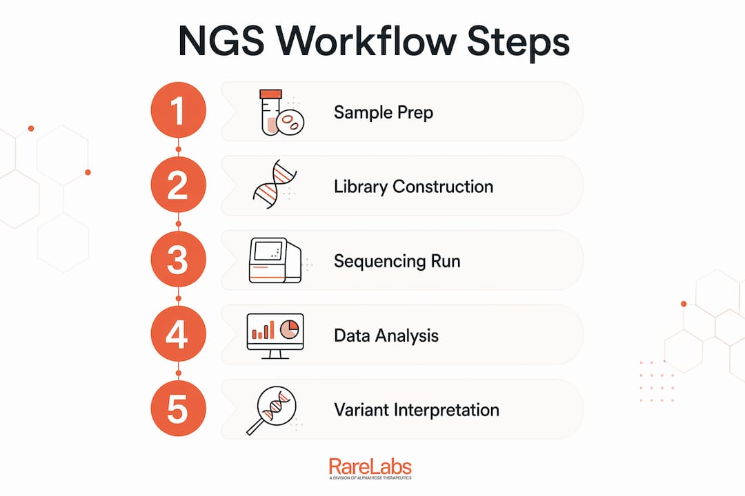
The standard NGS workflow: four steps where things go right or wrong
- Extraction. Isolate DNA or RNA from the sample. Quality here sets the ceiling for everything downstream. Degraded nucleic acids produce fragmented libraries with poor representation.
- Library preparation. Fragment the nucleic acids, ligate sequencing adapters, and quantify the result. Library preparation is among the most laborious steps and the most common source of run failure. Choices made here — PCR vs. PCR-free, fragmentation strategy, use of unique molecular identifiers (UMIs) — directly affect duplicate rates and allele-fraction accuracy.
- Sequencing. Loaded libraries run on the instrument. Chemistry varies by platform (sequencing-by-synthesis, semiconductor detection, single-molecule real-time, nanopore). The instrument outputs raw signal data converted to base calls.
- Bioinformatics. Primary analysis converts raw signal to FASTQ reads. Secondary analysis aligns reads to a reference genome and calls variants. Tertiary analysis annotates, filters, and interprets variants against clinical databases and ACMG criteria.
Pro Tip: Fluorometric quantification (e.g., Qubit) is more reliable than absorbance-based methods for library input. Inaccurate quantification is the single most common reason a library fails to cluster properly. Precise input measurement at the extraction and post-ligation steps pays dividends across every downstream stage.
Which sequencing platform fits your ultra-rare disease project?
Four platform families dominate current NGS work. Each has a distinct accuracy profile, read length, and clinical availability.

| Platform class | Read length | Accuracy / error profile | Throughput / cost profile | Best use cases | CLIA-validated assays available |
|---|---|---|---|---|---|
| Illumina (SBS, short-read) | — | high per-base accuracy; low substitution error | High throughput; lowest per-base cost | SNV/indel detection, panels, exomes, WGS, transcriptomics | Yes, widely |
| Thermo Fisher Scientific Ion Torrent (semiconductor, short-read) | — | High; homopolymer errors possible | Moderate throughput; benchtop-friendly | Targeted panels, oncology hotspots | Yes, selected assays |
| Pacific Biosciences PacBio (SMRT, long-read) | 10–25 kb average HiFi | high HiFi consensus accuracy | Lower throughput per run; higher per-base cost | Structural variants, repeat expansions, phasing, de novo assembly | Emerging |
| Oxford Nanopore Technologies (nanopore, long-read) | Up to hundreds of kb | ~99%+ with recent chemistry; higher raw error than SBS | Scalable from pocket to high-throughput; real-time output | Structural variants, methylation, rapid turnaround, direct RNA | Limited; growing |
Short-read platforms, particularly Illumina, remain the workhorse for clinical panels, exomes, and whole-genome sequencing (WGS) because of their low error rate and broad CLIA-validated assay availability. Long-read approaches from PacBio and Oxford Nanopore resolve structural variants, repeat expansions, and complex regions that short reads routinely miss — capabilities that matter considerably in ultra-rare disease cases where the causal variant is structural or sits in a low-complexity region.
- Choose short-read SBS for SNV/indel-heavy discovery and any result destined for clinical reporting.
- Choose long-read when prior short-read sequencing returned negative or ambiguous results, or when the phenotype suggests a structural or repeat-expansion etiology.
- Ion Torrent suits rapid targeted panels where benchtop footprint matters.
How clinical NGS differs from research NGS
NGS has moved firmly into clinical diagnostics, but the bar for a clinical-grade result is substantially higher than for research-grade data. The distinction matters when a variant call will influence a treatment decision or feed into a regulatory submission.
Clinical-grade NGS requires: validated assay performance (documented sensitivity and specificity by variant class), CLIA-accredited and CAP-inspected laboratory operations, ACMG-compliant variant classification (Pathogenic/Likely Pathogenic/VUS/Likely Benign/Benign), a structured clinical report with explicit coverage limitations, and documented pipeline versioning.
QA metrics to request from any lab:
- Mean coverage depth (target ≥100x for panels, ≥30x for WGS clinical, higher for somatic work)
- Base quality scores (Q30 percentage; >80% is a common threshold)
- Duplicate read rate (high duplicates indicate library complexity problems)
- On-target rate (percentage of reads mapping to the intended capture region)
- Uniformity (fraction of targets covered at ≥0.2x mean depth)
Research-grade sequencing without these validations is appropriate for model generation and hypothesis testing. It is not appropriate as the sole basis for a clinical treatment decision.
Pro Tip: Ask every testing lab for their pipeline name, version, reference genome build, and the validation study supporting their sensitivity/specificity claims by variant class. A lab that cannot provide this documentation is delivering research-grade data regardless of what the report header says.
How NGS supports discovery and treatment development for ultra-rare diseases
For undiagnosed cases, NGS enables discovery without prior sequence knowledge — the defining advantage over single-gene tests. A typical application flow looks like this: discovery sequencing (WGS or exome) → variant prioritization → patient-derived model generation (iPSC reprogramming, CRISPR editing) → functional screening → candidate therapeutic selection.
Specific application choices:
- Whole-genome or exome sequencing for cases with no clear candidate gene; exome covers a small portion of the genome but captures most known disease-causing variants at lower cost than WGS.
- Targeted gene panels when the phenotype maps to a defined gene set (e.g., a known channelopathy or metabolic pathway); faster turnaround, lower cost, higher coverage depth per target.
- RNA sequencing (RNAseq) to detect aberrant splicing, allele-specific expression, and fusion transcripts that DNA sequencing misses entirely. Particularly valuable when a variant of uncertain significance (VUS) needs functional evidence.
- Long-read sequencing for cases with suspected repeat expansions, structural rearrangements, or failed short-read resolution.
Sample type shapes what is possible. Blood is the standard for germline work. Fibroblasts from a skin punch biopsy are preferred for iPSC reprogramming. Biopsy tissue is used when the disease is tissue-specific. Minimum input requirements vary by platform and library type; confirm with the lab before collection.
Hopeatrarelabs connects NGS variant data directly to genetic disease modeling and parallel treatment screens, testing FDA-approved drugs, custom antisense oligonucleotides (ASOs), and gene therapy candidates against patient-derived models.
From raw reads to clinically useful variants: the bioinformatics pipeline
- Primary analysis. The instrument converts raw signal (fluorescence or ionic current) to base calls and outputs FASTQ files with per-base quality scores. Demultiplexing separates samples run in the same pool.
- Secondary analysis. An aligner (e.g., BWA-MEM for short reads) maps reads to the reference genome (GRCh38 is current standard). A variant caller (e.g., GATK HaplotypeCaller for germline, Mutect2 for somatic) identifies SNVs, indels, and copy number variants. Output: BAM/CRAM and VCF files.
- Tertiary analysis. Variants are annotated against databases (ClinVar, gnomAD, OMIM), filtered by frequency and predicted effect, and classified per ACMG criteria. This stage produces the clinical or research report.
Deliverables to require from any provider:
- Raw FASTQ files (retain for reanalysis)
- Aligned BAM or CRAM with index
- Annotated VCF with per-variant quality metrics (depth, allele fraction, genotype quality)
- Pipeline name, version, and reference genome build
- Annotation database names and versions
- Coverage summary per target region
- Documented sensitivity/specificity by variant class
Pro Tip: Retain raw FASTQ files and pipeline metadata. Annotation databases update continuously, and a VUS today may reclassify to Pathogenic within 18 months as new evidence accumulates. Reanalysis is only possible if the raw data and pipeline parameters are preserved.
Storage scale: a 30x WGS FASTQ pair runs roughly 90–120 GB; a BAM adds another 90 GB. Budget storage and compute accordingly for multi-patient projects.
Common NGS pitfalls and how to mitigate them
Short-read NGS misses structural variants larger than a few hundred base pairs, struggles with GC-extreme regions and repetitive sequences, and can produce false positives from PCR amplification artifacts or alignment errors. These are not edge cases; they affect real diagnostic yields.
- Poor input quality: Degraded DNA fragments below the library prep minimum produce low-complexity libraries. Use fluorometric QC and set a minimum DIN or RIN threshold before proceeding.
- PCR amplification bias: Duplicate reads inflate apparent coverage and distort allele fractions. PCR-free library preparation reduces this for WGS; UMIs help for low-input or somatic work.
- GC bias: Regions with extreme GC content are systematically under-represented. Spike-in controls and coverage QC reports reveal this before interpretation.
- Alignment artifacts: Reads mapping to paralogous regions generate false variant calls. Review BAM files in IGV for any clinically actionable call in a known segmental duplication.
- Structural variant blindness: Short reads cannot phase or span large rearrangements. Escalate to long-read or optical mapping when the phenotype is consistent with a structural etiology.
Orthogonal confirmation with Sanger sequencing or droplet digital PCR (ddPCR) remains standard practice before acting on a novel or clinically actionable variant. This is especially true for variants with low allele fraction or those sitting in difficult sequence contexts.
Pro Tip: A practical confirmation rule: any novel likely-pathogenic or pathogenic variant that will influence a treatment decision, and any variant with allele fraction below 20%, warrants orthogonal validation before clinical action. Multidisciplinary review — molecular geneticist, bioinformatician, and clinician together — catches interpretation errors that no single reviewer reliably catches alone.
How to choose between panel, exome, genome, RNAseq, and long-read sequencing
| Approach | Typical diagnostic yield context | Sample input | Turnaround | Cost shape | When to escalate |
|---|---|---|---|---|---|
| Targeted gene panel | Known phenotype, defined gene set | Low (ng range) | 2–4 weeks | Lowest | Negative result with strong phenotypic fit |
| Clinical exome (WES) | Unexplained Mendelian phenotype | Moderate | 4 weeks | Moderate | Negative exome + strong phenotype → WGS |
| Whole-genome (WGS) | Exome-negative, suspected structural/non-coding | Moderate–high | 6 weeks | Higher | Negative WGS → RNAseq or long-read |
| RNAseq | VUS needing functional evidence; splicing suspected | Tissue-specific | 3 weeks | Moderate | Complement to DNA sequencing, not replacement |
| Long-read (PacBio/ONT) | Repeat expansions, structural variants, phasing | Moderate–high | Variable | Higher per base | After short-read failure in structural variant context |
The practical decision rule: start with the smallest test likely to answer the question, then escalate if negative. A child with a well-defined metabolic phenotype and a known gene list starts with a panel. An unexplained neurodevelopmental disorder with no candidate gene goes straight to exome or WGS.
- Batching samples reduces per-sample cost on high-throughput runs; plan cohort sequencing when urgency allows.
- For urgent clinical situations (neonatal ICU, rapidly progressive disease), rapid WGS turnaround services exist and can return results in 24–72 hours at premium cost.
- Document the test scope, coverage limitations, and variant classes not detected in study protocols and patient consent forms. Patients and families deserve to know what the test cannot find.
Pro Tip: RNAseq from a disease-relevant tissue (not blood) adds functional evidence that resolves a substantial fraction of VUSs left ambiguous by DNA sequencing alone. If you have access to fibroblasts or iPSC-derived cells, RNAseq on those samples is often more informative than blood RNA for rare disease splicing questions.
Key Takeaways
NGS is the foundational technology for ultra-rare disease discovery, but its clinical value depends entirely on input quality, validated pipelines, and rigorous variant interpretation.
| Point | Details |
|---|---|
| Input quality is the rate-limiting step | Poor extraction or inaccurate quantification causes library failure before sequencing begins. |
| Platform choice follows the clinical question | Short-read SBS for SNV/indel work; long-read for structural variants, repeats, and phasing. |
| Clinical vs. research grade is a real distinction | CLIA/CAP accreditation, ACMG classification, and documented pipeline validation define clinical-grade results. |
| Orthogonal confirmation is non-negotiable | Novel or low-allele-fraction variants require Sanger or ddPCR confirmation before clinical action. |
| Hopeatrarelabs connects NGS findings to models | Variant data feeds directly into iPSC/CRISPR disease models and parallel treatment screens for ultra-rare cases. |
Why the bottleneck in NGS is never the sequencer
The field spent a decade celebrating falling sequencing costs, and rightly so. But the harder problem was always interpretation. A whole-genome run produces millions of variants; the clinical question is which one of them is causing this patient's disease. That question requires curated databases, validated pipelines, multidisciplinary expertise, and often functional evidence from a model system. Sequencing throughput solved itself. Interpretation infrastructure is still catching up.
For ultra-rare disease work specifically, the gap is even wider. Standard population databases have almost no carriers of the relevant variant. ACMG criteria were built around more common Mendelian conditions. The researcher or clinician working on a disease affecting fewer than a hundred known patients worldwide cannot simply look up the answer. They have to generate functional evidence, which means building a model, running a screen, and interpreting results in a disease context that no one has fully characterized before.
That is exactly where the combination of NGS and patient-derived modeling becomes something more than a workflow. It becomes the only realistic path to a hypothesis worth testing.
How Hopeatrarelabs translates NGS findings into patient-specific models
When a sequencing result identifies a candidate variant in a patient with an ultra-rare or undiagnosed disease, the next question is almost always: does this variant actually cause the phenotype, and can anything be done about it? Hopeatrarelabs is built to answer both.
Starting from an NGS variant call, Hopeatrarelabs generates patient-specific iPSC and CRISPR-edited disease models derived from the patient's own cells, then runs parallel treatment screens across thousands of FDA-approved drugs, custom ASOs, and gene therapy candidates. The work is translational research, not clinical diagnostics. It is designed for cases where no approved therapy exists and the standard pathway offers no near-term answer.
If you are a researcher, clinician, or family foundation working on an ultra-rare or undiagnosed genetic disease and have NGS data in hand, contact Hopeatrarelabs to discuss whether a personalized modeling and screening program fits your case.
Useful sources and further reading
- NCI Dictionary of Genetics Terms: Next-Generation Sequencing — Concise authoritative definition from the National Cancer Institute; good starting point for clinical communication.
- ACMG Standards and Guidelines for Variant Interpretation — The primary reference for variant classification in clinical NGS reporting; essential for any result entering a clinical workflow.
- CLIA and CAP Accreditation Information — Defines the regulatory requirements for clinical laboratory NGS testing in the United States.
- PMC: Next-Generation Sequencing Technology: Current Trends and Advancements — Peer-reviewed review covering platform comparisons, applications, and emerging directions; good for technical depth.
- PubMed: A Next-Generation Sequencing Primer for Clinicians — Accessible clinical primer covering interpretation challenges and the transition of NGS to diagnostic use.
- NCBI Bookshelf: Next-Generation Sequencing — Covers orthogonal validation requirements and clinical reporting standards; best for understanding confirmation practices.
- Illumina NGS Basics — Manufacturer-authored technical primer on SBS chemistry, workflow, and platform capabilities; useful for platform-specific technical detail.
- Thermo Fisher Scientific: What Is Next-Generation Sequencing? — Covers library preparation in detail, including common failure points; best for workflow troubleshooting context.