
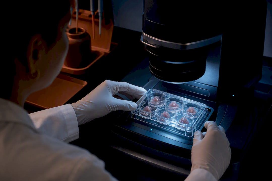
Patient-derived organoids, grown from either iPSCs or adult stem cells, give researchers a human-relevant, three-dimensional model system for studying disease mechanisms and running preclinical drug screens that animal models and flat cell cultures cannot replicate as faithfully. Paired with CRISPR-engineered isogenic controls, these systems now support drug repurposing, custom antisense oligonucleotide (ASO) testing, and gene therapy feasibility work for conditions with no existing treatment. The catch: reproducibility, missing vasculature, and incomplete tissue maturation still limit how far any single organoid experiment can extend before it needs validation elsewhere.
TL;DR:
- Reproducibility issues, lack of vascularization, and incomplete tissue maturation remain significant challenges for organoid experiments in rare disease models.
- Building organoids requires several weeks to months, with iPSC-derived models needing extra reprogramming steps and validation, which impacts clinical translation timelines.
- Isogenic controls created by CRISPR correction are essential to distinguish variant effects from genetic background noise, ensuring accurate cause-and-effect relationships.
- Organoid assays like the forskolin-induced swelling test in cystic fibrosis models are well-validated, whereas most other protocols still lack the same level of maturity and clinical correlation.
- Standardized protocols, multi-omics integration, and rigorous experimental design are critical for producing trustworthy data that can inform treatment decisions.
Table of Contents
- What Organoid Models Are and Why They Matter for Rare Genetic Disease Research
- How Do You Generate an Organoid Model From a Patient Sample?
- Building Genetic Controls: CRISPR, Isogenic Lines, and Multi-Omics
- Which Tissue Types Have the Strongest Organoid Evidence?
- How Are Organoids Used to Discover Rare Disease Treatments?
- Why Do Organoid Results Sometimes Fail to Reproduce?
- What Should a Rare Disease Organoid Study Protocol Include?
- Perspective: What Organoid Platforms Actually Change for Rare Disease Families
- Where RareLabs Fits Into an Organoid-Based Rare Disease Program
- Authoritative Reviews and Method Papers to Consult
- Sources
- FAQ
What Organoid Models Are and Why They Matter for Rare Genetic Disease Research
An organoid is a self-organized, three-dimensional structure grown from stem cells that mimics the architecture and function of a real organ, rather than a flat sheet of cells stuck to a plastic dish. For rare genetic disease research, that distinction is the whole point. A 2D fibroblast culture can tell you a gene is mutated. It cannot tell you how that mutation disrupts tissue-level signaling, cell-to-cell communication, or the physical gradients of oxygen and nutrients that shape real organ behavior. Organoid architecture allows those gradients to form, which is why organoids reproduce disease phenotypes that 2D systems routinely miss.
There are two main starting points for building a patient-specific organoid, and the choice shapes everything downstream.
- Induced pluripotent stem cells (iPSCs): reprogrammed from a patient's skin or blood cells, iPSCs can differentiate into nearly any tissue type, making them the default for neurological, muscular, and skeletal rare diseases where biopsy of the affected tissue is impractical or unsafe.
- Adult stem cells: harvested directly from a patient's biopsy (often intestinal crypts or airway epithelium), these retain some tissue memory and are the standard for epithelial organoids like the intestinal models used in cystic fibrosis research.
- Patient-derived vs. biopsy-derived: both approaches are "patient-derived" in the sense that they carry the patient's own genome, but iPSC lines require a reprogramming and differentiation step that adult stem cell organoids skip, trading a longer timeline for broader tissue access.
The tradeoff every lab has to accept is maturity. Most organoids, regardless of source, plateau at a fetal-like developmental state. A cerebral organoid rarely matches the synaptic complexity of an adult brain, and a cardiac organoid's contractile force typically falls short of a mature heart. For rare diseases that manifest in childhood or present with developmental phenotypes, that fetal-like state can actually be an advantage rather than a limitation. For late-onset or degenerative conditions, it is a real gap researchers need to plan around, usually with extended culture protocols or maturation cocktails.
Foundational reviews on advanced cellular models for rare disease study and broader analyses of 3D and organoid culture systems converge on the same conclusion: organoids sit in a middle zone between the simplicity of 2D culture and the cost, ethical burden, and species-specific biology of animal models. They will not replace either outright. They fill the gap where human genetic background and tissue architecture both matter, which describes most ultra-rare monogenic diseases fairly well. Hopeatrarelabs's own overview of how genetic disease modeling unlocks personalized rare care walks through how that translational rationale plays out for families dealing with undiagnosed conditions.
How Do You Generate an Organoid Model From a Patient Sample?
Building an organoid takes weeks to months, depending on tissue type, and every checkpoint along the way determines whether the final model is trustworthy enough to base a treatment decision on.
- Sample collection and reprogramming. A skin punch biopsy or blood draw provides the starting material for iPSC reprogramming, typically using Sendai virus or episomal vectors to avoid permanent genomic integration. Adult stem cell organoids skip this step and go straight from biopsy tissue into culture.
- Clonal expansion and banking. Multiple iPSC clones from the same patient are expanded and cryopreserved before differentiation begins. This step matters more than most labs initially budget for, because clonal variability is real even within a single patient's cells.
- Directed differentiation. Small-molecule cocktails and growth factors push the stem cells toward a target lineage, whether that is neural progenitor, cardiomyocyte, or intestinal epithelium. Timing and concentration here determine regional identity, which becomes critical for tissue-specific rare diseases.
- Organoid maturation. Cells self-organize into 3D structures over one to several weeks, often embedded in a scaffold that supports polarization and lumen formation.
- Quality control and validation. Before any experimental use, the organoid needs marker confirmation, functional assays, and, ideally, single-cell profiling to confirm it actually contains the cell types the protocol claims.
QC is where a lot of published organoid work quietly falls short. At minimum, a defensible model needs immunostaining for lineage-specific markers, single-cell RNA sequencing (scRNA-seq) to confirm cell-type proportions, and a functional readout appropriate to the tissue, electrophysiology for neurons, contractility measurements for cardiac or muscle organoids, or transepithelial resistance for epithelial models. Viability staining rounds out the panel, catching necrotic cores that form in larger organoids once diffusion limits kick in.
Pro Tip: Bank at least two to three independent iPSC clones per patient before you commit to a differentiation protocol. Clonal variation in differentiation efficiency is common enough that a single clone can give you a false negative on a phenotype that a sibling clone reproduces cleanly. Protocol notes on banking clones for patient-derived iPSC work cover the practical side of this in more depth.
Scaffold choice is its own decision point. Matrigel matrix and defined Collagen Type I remain the two dominant extracellular matrix (ECM) options, and they are not interchangeable. Matrigel is derived from mouse sarcoma cells and carries lot-to-lot variability that can quietly undermine reproducibility across a multi-year rare disease program. Defined synthetic or collagen-based matrices sacrifice some of Matrigel's biological richness for consistency, which matters more the longer a study runs. For a rare disease program that might span years and multiple funding cycles, that consistency argument usually wins.
Building Genetic Controls: CRISPR, Isogenic Lines, and Multi-Omics
The single biggest source of false conclusions in rare disease organoid work is comparing a patient's cells to an unrelated healthy donor's cells and attributing every difference to the disease variant. Genetic background varies enormously between unrelated individuals, and that noise can swamp a real but subtle phenotype.
CRISPR-Cas9 solves this by generating isogenic controls: the patient's own iPSC line, with the disease-causing variant corrected back to wild type, or a healthy donor line with the variant deliberately introduced. Integrating CRISPR with organoid platforms lets researchers validate that a specific mutation, not background genetic variation, drives the observed phenotype. This is the difference between "this patient's cells behave abnormally" and "this specific variant causes this specific abnormality," and only the second claim is useful for therapeutic development.
A well-designed genetic control strategy includes several layers:
- Correction and knock-in pairs: create both a corrected version of the patient line and, where feasible, a knock-in of the variant into an unrelated healthy background, so the phenotype shows up from two directions.
- Rescue experiments: reintroduce the wild-type gene or protein into the corrected line and confirm the phenotype reverses, which is stronger evidence than correction alone.
- Orthogonal functional readouts: never rely on a single assay. Pair a molecular readout (protein expression, calcium imaging) with a physiological one (contractility, electrophysiology, swelling assays).
- Blinded analysis: have a second team member score phenotypes without knowing genotype, particularly for morphological or imaging-based endpoints that are prone to observer bias.
Best-practice guidance on isogenic controls treats rescue experiments and orthogonal readouts as close to mandatory for any causality claim intended for publication or regulatory conversation, not optional extras.
Multi-omics integration adds the mechanistic depth that a single assay cannot. Layering transcriptomics, proteomics, and metabolomics onto the same organoid model, ideally the same physical sample split across platforms, surfaces pathway-level disruptions that a phenotypic assay alone would never flag. For an ultra-rare variant with no prior literature, that omics layer is often the only route to a testable hypothesis about mechanism, and it frequently surfaces candidate biomarkers that can later track treatment response in a living patient. An explainer on gene editing options for rare diseases covers how these correction strategies extend from research tools toward therapeutic candidates themselves.
Which Tissue Types Have the Strongest Organoid Evidence?
Organoid protocols are not one-size-fits-all, and the tissue you're modeling determines both the differentiation strategy and which functional readout will actually mean something.
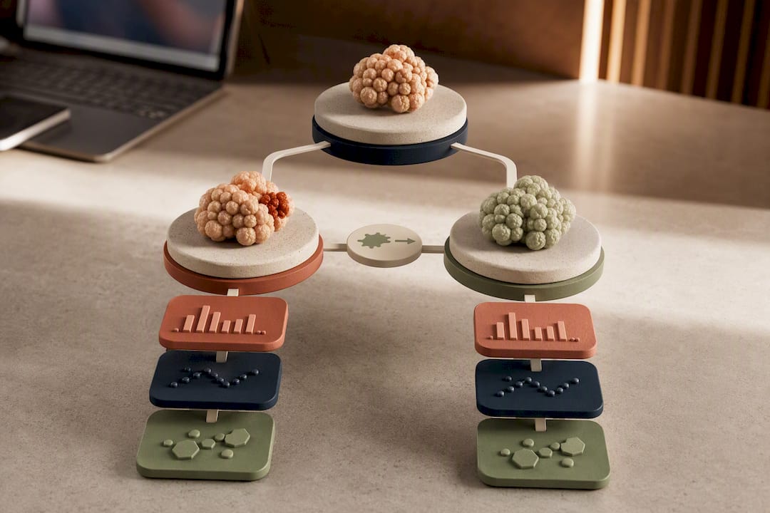
- Neural organoids: regionalization is not optional here. A generic cerebral organoid will miss disease biology that is specific to one brain region. Regionalized cerebellar organoids reproduced PCH2a-specific phenotypes that matched clinical imaging findings in patients, something a generalized neural organoid protocol would have missed entirely.
- Muscle and neuromuscular junction (NMJ) organoids: used for Duchenne muscular dystrophy (DMD) and related myopathies, these models pair muscle fiber differentiation with motor neuron co-culture to assess NMJ signaling defects functionally, not just structurally.
- Retinal organoids: gene-correction studies in RPGR-related retinitis pigmentosa have used patient-derived retinal organoids to confirm that CRISPR correction restores photoreceptor structure, a critical proof-of-concept step before any in vivo gene therapy trial.
- Intestinal and epithelial organoids: the forskolin-induced swelling (FIS) assay, which measures CFTR channel activity by tracking fluid-driven swelling in cystic fibrosis organoids, is one of the field's most mature phenotypic readouts and has been used directly to test gene-correction and small-molecule responses.
- Kidney and other epithelial organoids: increasingly used for nephrotoxicity prediction and modeling of rare renal genetic conditions where biopsy tissue is difficult to obtain repeatedly.
Organoid biobanks have shown that living collections of patient-derived organoids can correlate treatment response in the dish with actual clinical outcomes in several genetic disease and oncology studies, a signal that these models carry real predictive weight rather than being purely descriptive tools.
The FIS assay deserves particular attention because it demonstrates what a mature, validated organoid readout looks like once a field has had years to refine it. Cystic fibrosis organoid work has had roughly a decade head start over most other rare disease programs, and that maturity shows in how tightly FIS results track with clinical CFTR modulator response. Most other rare monogenic diseases are still building toward an assay with that level of validation, which is exactly the gap that regionalized, disease-specific protocol design is meant to close.
How Are Organoids Used to Discover Rare Disease Treatments?
The translational payoff of organoid modeling is running a treatment search on human tissue before ever touching a patient, and the workflow generally moves through three parallel tracks rather than one linear path.
High-throughput drug repurposing screens hundreds to thousands of FDA-approved compounds against a patient-derived organoid, looking for any existing drug that shifts the disease phenotype toward normal. Because these compounds already have human safety data, a hit can move toward off-label use or a formal trial far faster than a novel compound could. Hit triage matters as much as the screen itself: a phenotypic shift needs to be reproduced across multiple clones and, ideally, confirmed against an orthogonal readout before anyone treats it as a real signal. A practical guide to drug repurposing for rare diseases breaks down how that triage process typically runs.
Custom antisense oligonucleotide (ASO) testing takes a more targeted route, designing a short synthetic nucleotide sequence to correct splicing or knock down a toxic mRNA transcript specific to one patient's mutation. Organoids let researchers test multiple ASO candidates and dosing regimens against the actual disease phenotype before committing to the manufacturing and regulatory path required for a single-patient ASO program.
Gene therapy feasibility studies use organoids as the proving ground before any in vivo work. Testing a corrective CRISPR construct or a viral gene delivery vector in a patient's own organoid answers basic feasibility questions, does the correction actually restore function, is delivery efficient in this tissue type, cheaply and quickly relative to animal studies.
Translational consortia like ORGESTRA have used organoid workflows to accelerate personalized drug discovery for pediatric disorders including cystic fibrosis and cystinosis, and living biobanks built from these programs increase screening throughput while improving reproducibility across labs running the same rare variant. An overview of how disease modeling unlocks better rare disease treatments covers how these three tracks tend to run in parallel rather than sequentially for a single patient program.
Why Do Organoid Results Sometimes Fail to Reproduce?
Ask anyone who has run a multi-year organoid program what keeps them up at night, and reproducibility tops the list almost every time.
- Batch effects: different differentiation runs from the same iPSC line can produce organoids with meaningfully different cell-type proportions, especially past passage 20 or so.
- Incomplete maturation: fetal-like gene expression profiles persist in most protocols, limiting how well an organoid models adult-onset disease biology.
- Line-to-line heterogeneity: even isogenic pairs can drift genetically over extended culture, which is why periodic karyotyping and targeted sequencing checks matter.
- Protocol variability between labs: a differentiation protocol published in one paper rarely reproduces identically in another lab without extensive troubleshooting, a problem that has slowed cross-institution validation across the field.
Bioengineering is closing some of these gaps faster than protocol refinement alone ever could. Organoid-on-chip systems use microfluidic devices to deliver controlled perfusion, mimicking the nutrient and oxygen flow that a real vascular network would provide, which directly addresses the necrotic-core problem that limits how large a static organoid can grow before its center starts dying. Three-dimensional bioprinting adds spatial control that self-organization alone cannot achieve, letting researchers position different cell types in a defined architecture rather than hoping cells self-assemble into the right pattern. Advances in 3D bioprinting technique point toward digitally-guided deposition methods that could make organoid production far more consistent between batches and between labs.
Standardizing the ECM itself is the third leg of this effort. Moving from empirically-tuned natural hydrogels toward predictive material selection, choosing a matrix stiffness and composition based on known biology rather than trial and error, is a proposed remedy for the batch variability that plagues Matrigel-based protocols specifically.
Pro Tip: If your organoid program spans multiple years or funding cycles, lock your ECM lot and protocol version at the start and document every deviation. A change that seems minor, a different Matrigel lot, a two-degree temperature shift during differentiation, can shift phenotype penetrance enough to make year-three data incomparable to year-one data.
None of this fully satisfies what a regulatory review will eventually expect. Vascularization remains an open problem industry-wide, and no organoid protocol yet produces the kind of long-term, batch-independent consistency that a pivotal preclinical study ideally wants. That gap is closing, not closed.
What Should a Rare Disease Organoid Study Protocol Include?
A defensible organoid study, one that a physician, a foundation, or a regulatory reviewer can actually trust, follows a fairly predictable sequence of decisions made before any cells go into culture.
- Define regionalization and endpoints first. Decide which specific tissue region and which functional readout matter for this disease before starting differentiation, not after seeing preliminary results.
- Build isogenic controls alongside patient lines. Plan for CRISPR-corrected controls and, where possible, two to three independent patient clones from the outset.
- Predefine orthogonal assays. Commit to at least one molecular and one functional readout before the experiment starts, so results cannot be selectively reported after the fact.
- Plan multi-omics sampling. Reserve tissue for transcriptomic or proteomic profiling at the same timepoint as functional assays, not as an afterthought once a phenotype is confirmed.
- Implement blinded scoring. Assign phenotype scoring to someone who does not know genotype, especially for imaging or morphology-based endpoints.
- Standardize batch-correction methods. Decide in advance how differentiation batch will be tracked and statistically corrected for in the final analysis.
- Document full metadata. Record donor information, passage number, ECM lot, and protocol version for every organoid line generated.
| Checklist item | Why it matters | When to decide |
|---|---|---|
| Regionalization strategy | Wrong region misses tissue-specific pathology | Before differentiation begins |
| Isogenic controls | Isolates variant effect from genetic background | At line generation |
| Orthogonal readouts | Prevents single-assay false positives | Before first experiment |
| Blinded scoring | Removes observer bias | Before analysis |
| Metadata standards | Enables replication and biobanking | Continuously |
A step-by-step guide to genetic disease research walks through how this checklist maps onto a real program timeline for families and physicians scoping out an ultra-rare case.
Perspective: What Organoid Platforms Actually Change for Rare Disease Families
The honest case for organoid modeling isn't that it replaces animal studies or clinical trials. It's that it removes a specific kind of uncertainty that used to be unaddressable: what does this exact variant, in this exact patient's genetic background, actually do to human tissue? For decades, families with undiagnosed or ultra-rare conditions had no way to answer that question short of a clinical trial that would likely never happen, because no pharmaceutical company builds a trial for a disease affecting a handful of people worldwide.
Organoids change the economics of asking that question. A repurposing screen or an ASO feasibility test that once required years and millions of dollars in animal work can now run against a patient's own cells in months. That's not a cure delivery mechanism. It's a much faster, much cheaper way to rule out dead ends and identify the two or three candidates worth pursuing further.
Where I think the field oversells itself is in implying organoid data alone is translation-ready. It isn't, and treating a strong organoid phenotype as equivalent to clinical proof is a mistake that erodes trust when results don't replicate in patients. The realistic path forward runs through standardized data sharing, shared biobanks, and honest reporting of what an organoid model did and did not predict. Labs and biotech companies that build toward that standard, rather than around it, are the ones that will actually move rare disease therapeutics forward.
— John
Where RareLabs Fits Into an Organoid-Based Rare Disease Program
Everything in this article, from iPSC reprogramming to isogenic controls to repurposing screens, describes a workflow that most patients, families, and small research teams cannot execute without a dedicated lab partner. A lab can run a workflow built specifically for ultra-rare and undiagnosed genetic diseases, using a patient's own cells rather than a generic disease model that may not reflect their specific variant.
An engagement typically starts with a scoping conversation about the patient's genetic diagnosis, tissue type, and available biopsy or blood sample, followed by iPSC reprogramming and CRISPR-based isogenic control generation. From there, parallel treatment screens may test many FDA-approved drugs for repurposing candidates, develop custom ASOs where splicing correction is feasible, and evaluate gene therapy options where a viral or CRISPR-based correction makes sense. Results are compiled into a translational report intended for the patient's physician, giving the treating doctor something concrete to act on rather than a research paper with no clinical bridge.
Physicians, foundations, advocacy groups, and biopharma partners exploring a personalized discovery program for a specific rare disease can start by reviewing real workflow examples in the RareLabs Knowledge platform, which outlines how a program typically scopes, timelines, and what deliverables to expect before committing.
Authoritative Reviews and Method Papers to Consult
- Advanced cellular models for rare disease study covers neural, muscle, and skeletal organoid applications with disease-specific detail.
- Organoid genetic engineering and applications is the go-to reference for CRISPR integration and isogenic control design.
- 3D and organoid culture in research surveys technical limitations and engineering solutions across tissue types.
- Regionalized neural organoids for PCH2a is a concrete case study in why tissue regionalization matters.
- Corning's 3D organoid culture resource offers practical detail on ECM and scaffold selection.
This article is general information, not a substitute for advice from a qualified doctor. Consult a qualified healthcare professional about your own circumstances before acting on anything here.
Sources
- Advanced Cellular Models for Rare Disease Study: Exploring Neural, Muscle and Skeletal Organoids - PMC
- Organoid genetic engineering and applications - PMC
- 3D and organoid culture in research: physiology, hereditary genetic diseases and cancer - PMC
- Regionalized neural organoids for PCH2a - PMC
FAQ
What Are Organoid Models Used for in Rare Disease Research?
Organoid models let researchers study how a specific genetic variant disrupts human tissue architecture and function, and they serve as a preclinical screening platform for drug repurposing, ASO testing, and gene therapy feasibility before any patient trial.
How Long Does It Take to Build a Patient-Derived Organoid?
Generating and differentiating a patient-derived organoid typically takes several weeks to a few months, depending on tissue type, with iPSC-based protocols generally running longer than adult stem cell-derived organoids because of the added reprogramming step.
Why Are Isogenic Controls Necessary in Organoid Studies?
Isogenic controls, created through CRISPR correction of the patient's own cell line, isolate the effect of a specific disease variant from unrelated genetic background variation, which is essential for proving a mutation actually causes the observed phenotype.
What Are the Biggest Limitations of Organoid Models Today?
The main limitations are a lack of vasculatures causing necrotic cores in larger organoids, fetal-like maturation that limits modeling of adult-onset disease, and batch-to-batch variability that complicates reproducibility across labs.
Can Organoid Models Replace Animal Testing for Rare Diseases?
Organoid models reduce reliance on animal testing for early feasibility and mechanistic questions, but they do not fully replace in vivo validation, particularly for systemic effects, immune interactions, and long-term safety that isolated tissue models cannot capture.