
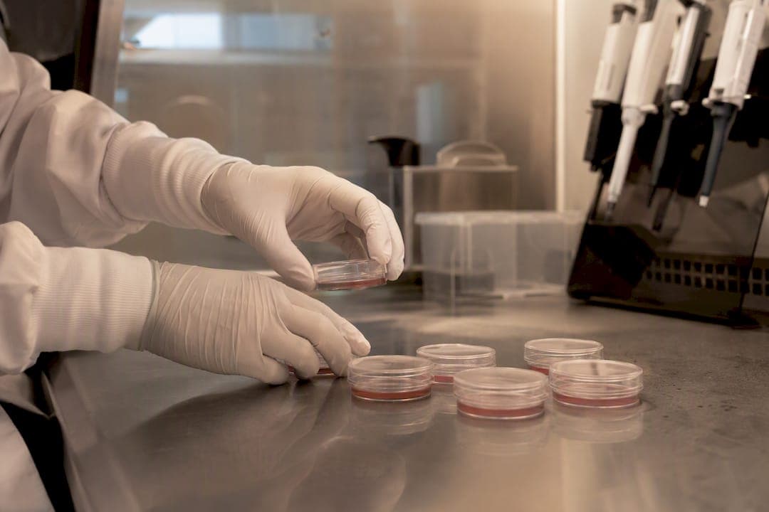
Patient-derived iPSCs are somatic cells, usually taken from skin or blood, reprogrammed to pluripotency while retaining the donor's exact genome. That genome retention is the whole point: it lets researchers build in vitro disease models, run precision drug screens, and generate translational data that animal models and immortalized cell lines can't replicate. The sections below walk through methods, model formats, assays, quality control, and where the science still runs into real limits.
TL;DR:
- Reprogramming from skin, blood, or urine depends on disease site and sample accessibility, with skin fibroblasts, blood, and urine as common sources.
- CRISPR-edited isogenic controls are essential for accurately attributing phenotypes to specific mutations, especially when comparing to different genetic backgrounds.
- Functional assays like multielectrode arrays and contractility tests provide network-level insights, but molecular profiling adds crucial orthogonal validation.
- Scaling production faces major challenges with labor-intensive clone selection, protocol variability, and GMP requirements, hindering large-cohort or therapeutic applications.
Table of Contents
- Why patient-derived iPSCs matter for disease research
- How are patient cells reprogrammed into iPSCs?
- Choosing between 2D, organoid, and organ-on-chip models
- When should you use CRISPR isogenic controls in iPSC studies?
- What assays work best with patient-derived iPSC models?
- How do you ensure quality control in iPSC research?
- From iPSC data to clinical decisions: what's the evidence bar?
- What's next for patient-derived iPSC research?
- How RareLabs applies patient-derived iPSCs in practice
- What ethical rules govern patient-derived iPSC research?
- How are patient-derived iPSC lines shared and biobanked?
- Why is scaling iPSC production still so hard?
- When should you choose patient-derived iPSCs over other models?
- Ready to start a personalized iPSC program?
- Sources
Why patient-derived iPSCs matter for disease research
The reason patient-derived iPSCs have taken over so much of translational biology is simple: they carry the donor's complete genetic background into a dish. Every risk allele, every modifier gene, every regulatory quirk that animal models can't reproduce comes along with the cell. That matters enormously for diseases where the phenotype depends on gene-gene interactions or genetic background effects that a knockout mouse simply doesn't have. Research on epilepsy modeling with patient-derived iPSCs shows these lines preserve disease-relevant effects that mouse models routinely miss, particularly for channelopathies and other electrophysiological disorders where species differences in ion channel biology distort the readout.
Compared with animal models, patient-derived iPSCs offer a human genomic context without cross-species extrapolation. Compared with immortalized cell lines (HeLa, HEK293, and the like), they offer disease relevance instead of a generic proliferative background that has drifted far from any normal tissue state. Neither alternative gives you a genuine window into how a specific patient's mutation behaves in human cells.
The core applications cluster into four buckets:
- Monogenic disease modeling — recapitulating single-gene disorders (cystic fibrosis, many epilepsies, Duchenne muscular dystrophy) where the causal variant is known and its cellular consequences can be directly observed.
- Complex disease modeling — capturing polygenic conditions (some cardiomyopathies, subsets of Parkinson's disease) where no single variant explains the phenotype, and only the patient's full genetic context reveals the mechanism.
- Cell therapy proof-of-concept — using differentiated iPSC progeny to test whether a corrected or engineered cell population can restore function before committing to a therapeutic program.
- Drug and toxicity screening — running compound panels against patient cells to find responders, non-responders, and off-target toxicity signals that generic cell lines can't flag.
None of this replaces animal studies outright. It fills a gap those models were never built to close: what happens in a human cell carrying this patient's exact mutation, at this dose, with this drug.
How are patient cells reprogrammed into iPSCs?
Reprogramming starts with a sampling decision, and that decision shapes everything downstream. Most programs pull from three somatic sources.
- Skin fibroblasts, obtained by a small punch biopsy, remain the classic source. They reprogram reliably and are well characterized, but the biopsy is invasive relative to a blood draw.
- Peripheral blood mononuclear cells (PBMCs) have become the default for many programs because a routine blood draw is far easier to arrange than a skin biopsy, especially for pediatric patients or those with mobility or bleeding concerns.
- Urine-derived cells offer a fully noninvasive option, useful when biopsy or blood draw isn't practical, though yields are lower and reprogramming efficiency varies more.
Tissue-of-origin matters more than most protocols acknowledge. For diseases with site-specific somatic mutations, reprogramming a skin fibroblast can miss the mutation entirely if it's confined to the affected organ. Liver disease modeling is the clearest example: work on patient-specific iPSCs for liver disease shows that deriving lines from diseased hepatic tissue, rather than an unrelated peripheral source, is sometimes the only way to capture the somatic mutations driving pathogenesis.
Reprogramming method is the second major fork. Non-integrating approaches, Sendai virus, episomal plasmids, and synthetic mRNA, have mostly displaced integrating retroviral and lentiviral methods because they don't leave residual transgene sequences in the genome that could confound later phenotyping. A methods paper generating an Alzheimer's disease iPSC line with an APP mutation used Sendai virus specifically because it preserves the disease mutation and produces a normal karyotype without genomic integration risk. Episomal reprogramming is cheaper and avoids viral handling, but efficiency runs lower and typically demands more colony picking. mRNA reprogramming is fast but technically fussier, requiring repeated transfections over roughly two weeks.
The operational workflow, once you have institutional approval, typically runs: consent and IRB clearance, sample collection and cold-chain transport to the lab, reprogramming (2 to 4 weeks depending on method), initial colony picking, and a first pluripotency screen before banking. Expect 6 to 10 weeks from consent to a banked, characterized line, longer if the sample needs multiple reprogramming attempts.
Pro Tip: Bank at least two to three independent clones per patient from the start. Clonal variation in reprogramming efficiency and residual epigenetic memory is common, and having backup clones saves you from restarting the entire derivation if your primary clone fails karyotype or pluripotency QC months later.
Choosing between 2D, organoid, and organ-on-chip models
Once a line is banked and characterized, the next decision is differentiation format, and this is where a lot of programs either overspend on complexity they don't need or underinvest in physiological realism they actually require.
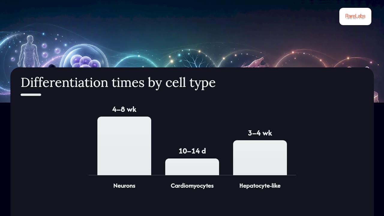
Directed differentiation protocols push iPSCs toward specific lineages using staged growth factor and small-molecule cocktails. Neurons typically take 4 to 8 weeks to reach a usable functional state, depending on subtype. Cardiomyocyte differentiation protocols using modulation of the Wnt pathway can produce beating cardiomyocytes within 10 to 14 days, though full electrophysiological and metabolic maturity takes considerably longer. Hepatocyte-like cells generally require 3 to 4 weeks and often remain functionally immature compared with adult liver tissue, a persistent limitation across the field.
Model format then becomes a question of what you actually need to answer:
- 2D monolayers are simple, cheap, and scale well for throughput. They're the right call for compound screening and basic mechanistic questions where cell-autonomous behavior is what matters.
- 3D organoids better replicate tissue architecture and cell-cell interactions. A review of iPSC model formats documents the field's shift toward organoids and multi-organ "body-on-chip" systems specifically because 2D culture flattens out interactions that drive real tissue behavior.
- Organ-on-chip systems add physiologic fluid flow and, increasingly, connections between multiple organ modules, letting researchers study drug metabolism and toxicity (ADMET) across organ systems rather than in isolation.
As a selection guide: if the question is "does this compound hit this target," 2D screening is faster and cheaper. If the question is "how does this tissue actually behave structurally," organoids earn their added cost and complexity. If the question involves systemic drug behavior, absorption, distribution, metabolism, excretion, across connected organ systems, only a multi-organ chip gives you that data. Matching format to question, rather than defaulting to whichever model is trendiest, is what keeps a program on budget and on timeline.
When should you use CRISPR isogenic controls in iPSC studies?
Patient-derived lines have one persistent weakness: every patient carries a unique genetic background, so comparing patient cells to a different, unrelated healthy donor line always leaves open the question of whether an observed phenotype comes from the disease mutation or just from normal genetic variation between two different people. CRISPR-generated isogenic controls solve this by editing the mutation directly into or out of the same genetic background.
Three isogenic strategies cover most use cases:
- Correction — repairing the patient's mutation back to the wild-type sequence in their own cell line, isolating the mutation's effect against an otherwise identical genome.
- Knock-in — introducing the patient's specific variant into a healthy control line, useful when the patient's own cells are hard to reprogram or maintain.
- Revertant — reverting an edited or corrected line back to the original mutant state, a useful cross-check that the correction itself, not an off-target edit, drove the observed change.
An Annual Reviews analysis of iPSC disease biology treats CRISPR-derived isogenic lines as essential for causal genotype-phenotype inference precisely because they remove genetic background as a confounder. Combining patient-derived collections with isogenic engineering, rather than choosing one over the other, is what JAX's comparison of engineered versus patient-derived iPSCs identifies as the strongest design for both reproducibility and patient-specific insight.
Mitigating confounders further requires testing multiple independent clones per edit (single-clone conclusions are a common failure mode), running off-target sequencing on any edited line, and validating key findings with an orthogonal assay rather than relying on one readout alone.
Recommended QC for edited lines before you trust any downstream data: long-range PCR across the edit site to confirm the intended change and rule out unintended insertions, SNP or CNV array analysis to catch larger structural changes CRISPR can introduce, and a standard karyotype to confirm no gross chromosomal abnormality emerged during editing and clonal expansion.
Pro Tip: Run your off-target analysis before you invest in downstream phenotyping, not after. Catching a problematic edit at the QC stage costs you a week; catching it after six months of functional assays costs you the whole dataset. Our CRISPR technology guide covers editing options and control designs in more depth.
What assays work best with patient-derived iPSC models?
Assay choice depends on what kind of signal you're chasing, electrical, mechanical, molecular, and how many conditions you need to run in parallel.
Functional platforms dominate early-stage phenotyping. Multielectrode arrays (MEA) capture electrophysiological activity from iPSC-derived neurons or cardiomyocytes at the network level, and they've produced some of the field's clearest translational wins. The epilepsy research cited earlier describes MEA screening of patient-derived neurons identifying compounds that moved forward into clinical evaluation, a rare and concrete example of iPSC data directly informing a treatment decision. Contractility assays measure force generation in iPSC-derived cardiomyocytes, critical for cardiotoxicity screening. Calcium imaging tracks intracellular signaling dynamics and works well alongside either MEA or contractility readouts as a complementary signal.
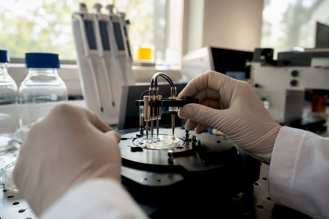
High-throughput screening design matters as much as the assay itself. Standard 384- or 1536-well plate formats let you screen sizable compound libraries, but only if you build in proper positive and negative controls on every plate to catch plate-to-plate drift. Screening FDA-approved compound libraries for repurposing is increasingly common in rare disease work, since these compounds already carry human safety data, which can shorten the path from a screening hit to a feasible next step. Our guide on prioritizing drug candidates using patient iPSCs walks through practical hit-ranking criteria.
Molecular phenotyping rounds out the picture as an orthogonal validator rather than a primary screen. RNA-seq catches transcriptional signatures a functional assay might miss entirely. Single-cell profiling reveals population heterogeneity that bulk assays average away, often the difference between "no effect" and "effect in a specific subpopulation." Proteomics adds a layer functional assays can't: confirming that a transcriptional change actually produces a protein-level consequence.
How do you ensure quality control in iPSC research?
Reprogramming and differentiation both introduce risk of genomic instability, and a review of iPSC model development flags genomic instability and persistent epigenetic memory from the donor tissue as recurring issues that undermine reproducibility if left unchecked. A working QC checklist should never skip these steps.
- Pluripotency marker panels — confirm expression of core markers (OCT4, SOX2, NANOG, and surface markers like TRA-1-60) by flow cytometry or immunostaining before any line moves into differentiation.
- Mycoplasma testing — run this on every new line and periodically thereafter; contamination silently distorts phenotyping data for months before anyone notices.
- Karyotype and genomic stability testing — standard karyotyping plus, where budget allows, SNP array analysis to catch subchromosomal changes that a karyotype alone would miss.
- Replication design — build studies around multiple independent clones per patient, true biological replicates (not just technical repeats of the same well), and independent differentiation runs performed on separate days.
Common artifacts to watch for: incomplete silencing of reprogramming factors (check by testing for residual vector sequences if you used an integrating method), differentiation efficiency that drifts between batches of the same protocol, and epigenetic memory where a line retains marks from its tissue of origin that bias it toward or away from certain differentiation paths. Detecting these usually means running a small pilot differentiation on every new line before committing it to a full study, rather than assuming a passed pluripotency check guarantees clean downstream behavior.
From iPSC data to clinical decisions: what's the evidence bar?
Patient-derived iPSC data feeds into translational decisions in two main ways. The first is drug repurposing: screening patient cells against FDA-approved compound libraries to find candidates whose mechanism plausibly addresses the patient's specific mutation, then using that signal to prioritize which existing drug is worth a compassionate-use or expanded-access attempt. The second is preclinical trial design: using iPSC-derived phenotypic and dose-response data to inform endpoints, dosing ranges, or patient stratification criteria before an early-phase trial ever starts.
Cell therapy applications carry a distinct regulatory bar. Any iPSC-derived cell product intended for therapeutic use faces scrutiny on three fronts:
- Potency — demonstrating the differentiated product actually performs the intended function at a consistent, measurable level batch to batch.
- Tumorigenicity — confirming no residual undifferentiated pluripotent cells remain, since even a small contaminating population carries teratoma risk.
- GMP-compliant derivation — the entire reprogramming and differentiation process needs to meet Good Manufacturing Practice standards if the product will ever be administered to a patient, which is a substantially higher bar than research-grade derivation.
Presenting iPSC-derived evidence to clinicians and regulators requires more rigor than an academic paper typically demands. Replication across independent clones and independent differentiation runs is table stakes, not optional. Orthogonal validation, confirming a functional finding with an independent molecular readout, strengthens a case considerably more than repeating the same assay. Effect size reporting matters as much as statistical significance: a regulator or treating physician needs to know whether an observed drug response is large enough to plausibly matter clinically, not just whether a p-value cleared a threshold. Our piece on regenerative medicine applications of pluripotent stem cells covers how these standards apply once a program moves toward cell therapy development.
What's next for patient-derived iPSC research?
The biggest near-term bottleneck isn't the biology, it's standardization. Cohort studies and high-throughput pipelines both demand consistent, comparable lines across labs and patients, and right now protocol variation between labs remains a real source of noise in cross-study comparisons.
Maturation is the second persistent problem. iPSC-derived cardiomyocytes, neurons, and hepatocytes routinely fall short of full adult functional maturity, which limits how confidently findings translate to actual patient physiology. Bioengineering approaches, mechanical loading for cardiomyocytes, extended culture with metabolic switching cues, co-culture with supporting cell types, are closing that gap incrementally rather than solving it outright. Multi-organ integration, connecting differentiated modules on a single chip, is the most promising route toward physiologically realistic systemic modeling.
Three developments are worth watching over the next several years:
- Single-cell phenotyping at scale — resolving population heterogeneity within a single patient's cells, rather than treating a differentiated culture as a uniform population.
- AI-assisted phenotype discovery — using pattern recognition across large imaging and molecular datasets to flag subtle phenotypic differences a human reviewer would likely miss.
- Patient-cohort iPSC consortia — pooled biobanks and shared protocols across institutions that increase statistical power for rare conditions where any single lab's patient pool is too small to draw firm conclusions alone.
None of these close the maturation gap overnight, but together they're shrinking the distance between an interesting dish finding and a decision a physician can actually act on.
How RareLabs applies patient-derived iPSCs in practice
Hopeatrarelabs builds personalized disease models directly from a patient's own cells: iPSC derivation, CRISPR-generated isogenic controls to isolate the mutation's effect, and high-throughput screening against FDA-approved drugs and custom antisense oligonucleotides (ASOs). The goal in every program is the same: turn a patient's genome into a testable model fast enough to matter for a family still searching for options.
- Derivation and characterization of patient-specific iPSC lines from accessible tissue sources
- CRISPR-edited isogenic controls to confirm a mutation, not genetic background, drives the observed phenotype
- Parallel screening across thousands of repurposed drug candidates and custom ASO designs
What ethical rules govern patient-derived iPSC research?
Deriving iPSCs from a patient's cells starts with informed consent, and that consent has to cover more ground than a standard biopsy release. Patients and families need to understand that their cells will be reprogrammed, potentially genetically edited, banked long-term, and possibly shared with collaborating labs or biobanks, sometimes years after the original sample was collected.
Consent protocols typically address a few distinct issues at once: whether the line can be used beyond the original study, whether it can be shared with outside researchers or commercial partners, whether incidental genetic findings unrelated to the original disease question will be returned to the patient, and how long the line will be retained. Pediatric consent adds another layer, since a minor's cells are often collected under parental consent for a condition that may not yet have a clear diagnosis, which makes broad future-use language especially important to get right at the outset.
Institutional Review Board (IRB) oversight governs all of this, and any program working with human-derived cell lines needs IRB approval before sample collection begins, not after. For rare and undiagnosed disease families in particular, consent conversations often carry real emotional weight: families are frequently consenting to research with no guarantee of a therapeutic outcome for their own child, which makes clear, honest communication about realistic timelines and probabilities part of the ethical obligation, not just a legal formality.
How are patient-derived iPSC lines shared and biobanked?
Once a line clears pluripotency and genomic stability QC, the next question is where it lives long-term and who else can use it. Biobanking patient-derived iPSC lines allows a single successful derivation to support multiple studies over years, which matters enormously for rare diseases where recruiting a second patient with the same mutation may simply not be possible.
Data sharing around these lines typically includes the genomic and phenotypic characterization data alongside the physical cell line itself: karyotype results, pluripotency marker panels, whole-genome or targeted sequencing confirming the disease variant, and differentiation efficiency data from prior studies. Sharing that metadata is often more valuable to the broader research community than the cells alone, since it lets other labs judge whether a given line fits their own study design before requesting it.
Consent scope directly determines what a biobank can do with a line. A line consented only for a single named study cannot legally be redistributed to outside collaborators, no matter how scientifically valuable it would be to a broader consortium. This is precisely why broad, future-use consent language, gathered honestly and transparently at the point of collection, has become such a priority for rare disease programs: it's the difference between a line that helps one study and a line that helps every family with that same mutation for years afterward. Coordination across institutional biobanks, rather than each lab hoarding its own lines, is the direction the field is slowly moving, though formal cross-institution data-sharing standards remain a work in progress.
Why is scaling iPSC production still so hard?
Producing a single well-characterized iPSC line for one research study is a solved problem. Producing hundreds of lines, consistently, for a cohort study or a therapeutic screening pipeline, is not, and the gap between those two scales is where most programs hit friction.
Manual colony picking and clone selection, still standard in many labs, simply doesn't scale past a handful of lines without proportional increases in skilled labor. Batch-to-batch variability in reprogramming efficiency means some patient samples yield usable lines quickly while others require multiple attempts, which makes timeline planning for large cohorts genuinely difficult. Differentiation adds a second scaling bottleneck: protocols optimized in a research lab on a small scale often behave differently when pushed toward higher-throughput formats, requiring re-optimization rather than simple duplication.
For therapeutic applications specifically, GMP-compliant production multiplies the difficulty. Research-grade derivation and GMP derivation are not the same process scaled up. They require different facilities, different reagent sourcing (research reagents versus clinical-grade), and different documentation at every step, which is a major reason so few iPSC-derived cell therapies have made it past early-phase trials despite years of promising preclinical data. Automation, closed-system bioreactors, and standardized protocols across labs are the main levers being developed to close this gap, but for now, scaling remains one of the slowest, most expensive parts of moving from a promising dish finding to something a patient can actually receive.
When should you choose patient-derived iPSCs over other models?
Choose patient-derived iPSCs when the question depends on a specific patient's genetic background, drug response prediction, mechanism in a monogenic disease, feasibility of a personalized therapy. Choose CRISPR isogenic lines when you need to isolate a single variant's causal effect against a clean genetic background. Choose animal models when you need whole-organism physiology, immune interaction, or long-term in vivo safety data that no dish-based system can provide.
The strongest designs rarely rely on just one. Pairing a patient-derived line with its isogenic-corrected counterpart, then validating the key finding in an animal model before clinical translation, gives you patient specificity, causal certainty, and systemic context in one program.
Pro Tip: When presenting iPSC findings to a translational partner or funder, lead with the isogenic comparison, not the raw patient-versus-healthy-donor result. It preempts the first question every skeptical reviewer asks: how do you know it's the mutation and not just genetic noise?
— John
Ready to start a personalized iPSC program?
If you've been piecing together disease modeling, drug screening, and genetic controls across separate vendors, Hopeatrarelabs collapses that into one coordinated program instead of three disconnected contracts. Patients, families, foundations, physicians, and biopharma partners come to Hopeatrarelabs because it runs derivation, CRISPR isogenic control generation, and parallel screening against thousands of FDA-approved drugs and custom ASOs under a single roof, which matters most when a rare disease timeline doesn't leave room for handoffs between separate labs.
Every program starts with an exploratory consultation to assess whether a patient's specific mutation and clinical history make a personalized modeling program feasible. From there, Hopeatrarelabs outlines a derivation and screening plan built around that patient's genome, not a generic protocol adapted after the fact. If you're a patient, family, physician, or research partner ready to find out what a personalized program could look like for your specific case, start a program with RareLabs to request that first consultation.
This article is general information, not a substitute for advice from a qualified doctor. Consult a qualified healthcare professional about your own circumstances before acting on anything here.
Sources
- Using Patient-Derived Induced Pluripotent Stem Cells to Model and Treat Epilepsies
- Induced Pluripotent Stem Cells in Disease Biology and the Evidence for Their In Vitro Utility | Annual Reviews