
Duchenne muscular dystrophy was first described clinically in the 1860s by Guillaume-Benjamin Amand Duchenne, though earlier brief reports appeared in the 1830s. The molecular story came more than a century later: the DMD gene was identified in 1986 through positional cloning, and the protein it encodes, dystrophin, was characterized and named in 1987. Those are two genuinely different discoveries. The 19th-century work told physicians what the disease looked like; the 1980s work told scientists why it happens and, critically, how to target it.
Key Takeaways
The DMD gene was identified in 1986 through positional cloning, dystrophin was named in 1987, and every mutation-specific therapy developed since traces its design logic directly to those two molecular milestones.
| Point | Details |
|---|---|
| Clinical description: 1860s | Duchenne's biopsy and photographic work (1861–1868) established the foundational clinical picture of DMD. |
| Earlier reports: 1830s | Semmola (1834) and Conte (1836) published brief descriptions, but neither gained wide recognition. |
| DMD gene identified: 1986 | Positional cloning by Kunkel, Monaco, and Koenig located and cloned the DMD gene at Xp21. |
| Dystrophin named: 1987 | Hoffman, Koenig, and Kunkel published the full cDNA sequence and confirmed dystrophin's absence in DMD muscle. |
| Practical consequence | Mutation-specific diagnostics, ASO therapies, gene therapy vectors, and iPSC models all depend on the 1987 gene/protein knowledge. |
Table of Contents
- When was DMD first described, and by whom?
- How the DMD gene and dystrophin were identified in the 1980s
- How the gene discovery changed diagnosis and opened therapy pathways
- Landmark papers and researchers worth reading
- Why the 1986–1987 discovery still drives modern patient-specific research
- What the DMD timeline teaches us about rare disease research
- Sources
When was DMD first described, and by whom?
The earliest documented descriptions of a condition matching DMD appear in Italian medical literature. Giovanni Semmola reported a case in 1834, and Gaetano Conte followed with a more detailed account in 1836. Neither report gained wide circulation, and neither physician connected the clinical picture to a distinct disease entity.
Guillaume-Benjamin Amand Duchenne de Boulogne changed that. Working in Paris through the 1860s, Duchenne combined systematic clinical observation with two tools that were genuinely novel for the era: muscle biopsy and medical photography. His biopsy work and photographic records from 1861 to 1868 produced the most complete clinical portrait of the disease that existed for decades, which is why it carries his name. He documented the hallmark pattern of progressive proximal weakness beginning in early childhood, the characteristic calf enlargement (pseudo-hypertrophy caused by fat and connective tissue replacing muscle), and the relentless course that left boys wheelchair-bound and typically caused death in early adulthood.
William Richard Gowers extended that clinical foundation in 1879, describing the now-famous Gowers' sign: the way affected boys push themselves upright from the floor using their arms against their own legs, a compensatory maneuver that reflects severe hip and thigh weakness. Gowers also contributed careful natural history data that helped distinguish DMD from other forms of muscular weakness.
Early clinical contributors at a glance:
- Giovanni Semmola (1834): First documented case report consistent with DMD
- Gaetano Conte (1836): More detailed early Italian description
- Guillaume Duchenne (1861–1868): Systematic biopsy-based clinical characterization; gave the disease its name
- William Gowers (1879): Described Gowers' sign; contributed natural history data
- Edward Meryon (1852): British physician who noted the hereditary pattern and distinguished the condition from spinal disease
For roughly 120 years after Duchenne's work, the disease was understood entirely through its clinical presentation. Physicians could recognize it, describe its progression, and document its inheritance pattern. What they could not do was explain the underlying defect or intervene at its root cause.
How the DMD gene and dystrophin were identified in the 1980s
The molecular breakthrough came through a strategy called positional cloning, sometimes called reverse genetics. The approach works backward from a chromosomal location to the gene itself, without needing to know the protein first. For DMD, researchers knew from X-linked inheritance patterns that the gene sat on the X chromosome. The challenge was finding it within a genome that, at the time, had no complete map.
OMIM describes the study of DMD as one of the earliest notable successes of positional cloning in humans, a precedent-setting achievement that demonstrated the strategy could work for diseases where the protein was completely unknown. That precedent mattered: it opened the door for dozens of other disease-gene discoveries in the years that followed.
The key events unfolded rapidly between 1985 and 1987:
| Year | Milestone | Key Investigators |
|---|---|---|
| 1985 | Chromosomal locus narrowed to Xp21 using deletion mapping in affected males | Louis M. Kunkel, Anthony Monaco |
| 1986 | First fragments of DMD gene cDNA identified; gene region cloned | Michel Koenig, Louis M. Kunkel, Anthony Monaco |
| 1986 | Public announcement of DMD gene identification | Kunkel lab (Harvard) |
| 1987 | Full cDNA cloned; predicted protein sequence published; protein named dystrophin | Michel Koenig, Eric P. Hoffman, Louis M. Kunkel |
| 1987 | Antibody-based detection confirmed dystrophin absent in DMD muscle, present in normal muscle | Eric P. Hoffman and colleagues |
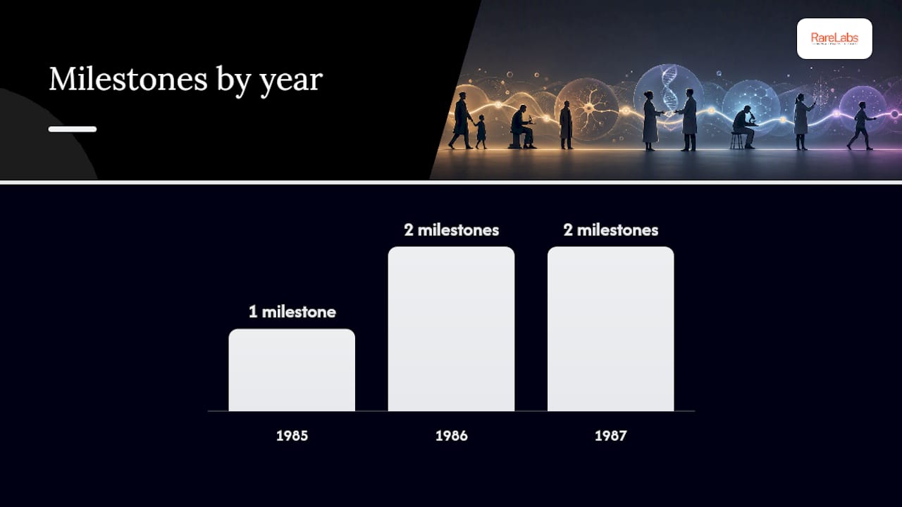
The first cDNA fragments were identified in 1986, with the predicted protein sequence and antibody-based detection following in 1987, enabling the identification and naming of dystrophin. The protein was very large, making it one of the largest proteins encoded in the human genome. Immunoblot experiments showed it clearly present in healthy muscle and absent in muscle from DMD patients, a clean result that confirmed the gene-to-disease connection.
The principal investigators and their roles:
- Louis M. Kunkel (Harvard): Led the positional cloning effort; his lab identified the chromosomal region and drove the cDNA cloning race
- Anthony Monaco: Key collaborator in the Kunkel lab during the Xp21 mapping and early cloning work
- Michel Koenig: Central to the full cDNA cloning and the predicted protein sequence published in 1987
- Eric P. Hoffman: Performed the antibody-based experiments that confirmed dystrophin's absence in DMD tissue and its presence in normal muscle; co-named the protein
The MDA's milestones document records Lou Kunkel's team among the key contributors to the mid-1980s mapping and cloning work, with dystrophin fully characterized by 1987.
How the gene discovery changed diagnosis and opened therapy pathways
Before 1986, diagnosing DMD meant combining clinical findings with elevated creatine kinase levels and muscle biopsy results. Carrier testing was probabilistic at best. After the gene was cloned and dystrophin identified, the entire diagnostic framework shifted.
The MDA notes that identifying the DMD gene in 1986 and the dystrophin protein in 1987 clarified disease mechanism and paved the way for mutation-specific diagnostics. Genetic sequencing could now pinpoint the exact mutation in a given patient, whether a large deletion, a duplication, or a point mutation. That precision matters clinically because different mutation types respond to different therapeutic strategies.
The therapy pathways unlocked by the discovery include:
- Antisense oligonucleotides (ASOs): Designed to skip specific exons and restore a partial reading frame; only feasible once the gene sequence and mutation landscape were known
- Gene therapy vectors: AAV-based approaches delivering a truncated but functional dystrophin (micro-dystrophin) depend entirely on knowing the protein's functional domains
- CRISPR-based correction: Editing out or correcting specific mutations requires exact knowledge of the gene sequence
- Improved animal models: The mdx mouse was confirmed as a dystrophin-deficient model only after the protein was identified, giving researchers a validated preclinical platform
- Carrier testing and prenatal diagnosis: Standardized DNA-based testing replaced probabilistic enzyme assays for families with a known mutation
Pro Tip: If you or a family member has a DMD diagnosis, knowing the precise mutation type, not just the clinical diagnosis, determines eligibility for mutation-specific trials and approved exon-skipping therapies. Ask your neurologist for full DMD gene sequencing if only a clinical or biopsy diagnosis exists. A gene variant interpretation guide can help you understand what your sequencing report means.
The 1986–1987 discovery established the practical framework for mutation-specific diagnostics and therapies; without positional cloning and dystrophin identification, ASO and gene therapy strategies would not be feasible. That is not an overstatement. Every FDA-approved exon-skipping drug for DMD traces its design logic directly back to the 1987 cDNA sequence.
Landmark papers and researchers worth reading
The primary literature from this period is dense but accessible to anyone willing to work through the methods sections. Here is a compact reading list with context for each entry:
-
Koenig et al. (1987), Cell: The paper that published the complete DMD cDNA sequence and predicted the protein. This is the foundational molecular paper. Look for the reading frame analysis and the predicted amino acid sequence, which established dystrophin as a structural protein related to spectrin.
-
Hoffman, Brown, and Kunkel (1987), Cell: The paper that named dystrophin and showed its absence in DMD muscle using antibodies. The immunoblot figures are the clearest demonstration of the gene-to-protein-to-disease connection. Hoffman's antibody work made the discovery tangible for clinicians.
-
Monaco et al. (1986), Nature: The cloning paper that identified the first fragments of the DMD gene. Read the methods for an illustration of how positional cloning worked before the human genome was sequenced.
-
PMC review on the discovery of dystrophin: A retrospective that places the 1986–1987 papers in historical context. Good starting point before reading the primary papers; it explains what was known before the cloning and what changed afterward.
-
OMIM entry 300377: The curated genetic database entry for the DMD gene. Updated regularly; useful for tracking how the mutation catalog has grown since 1987 and for understanding genotype-phenotype correlations.
Key researchers to follow in the primary literature:
- Louis M. Kunkel: Search PubMed for his 1985–1988 papers to trace the positional cloning strategy from chromosomal mapping to full gene identification
- Eric P. Hoffman: His 1987 Cell paper is the clearest entry point; his subsequent work on dystrophin-associated proteins extended the molecular picture considerably
- Michel Koenig: Central to the cDNA sequencing work; his papers detail the reading frame and protein structure predictions
- Anthony Monaco: Key contributor to the early Xp21 mapping work that made the cloning race possible
When reading primary papers from this era, pay attention to sample size (often small by modern standards), the methods used to confirm protein identity (antibody specificity matters), and whether findings were replicated in independent patient cohorts. The 1987 papers hold up well on all three counts.
Why the 1986–1987 discovery still drives modern patient-specific research
Knowing the DMD gene sequence did not just explain the disease. It created the infrastructure for every meaningful research advance that followed. The mdx mouse, confirmed as a dystrophin-deficient model after the protein's identification, gave researchers a validated in vivo system that accelerated preclinical testing. Human iPSC models came later, but they depend on the same gene-level knowledge: you cannot model a specific DMD deletion in a stem cell without knowing exactly where that deletion sits in the 2.4-megabase gene.
Modern methods that rely directly on the 1987 gene and protein knowledge:
- iPSC disease modeling: Patient-derived induced pluripotent stem cells are differentiated into cardiomyocytes or skeletal muscle cells carrying the patient's exact DMD mutation, producing a living cellular model of the disease
- CRISPR isogenic controls: A patient iPSC line is corrected at the mutation site to create a genetically identical "healthy" control, isolating the mutation's effect from genetic background noise
- ASO design: Antisense oligonucleotides are designed to target specific exon-intron boundaries defined by the 1987 cDNA sequence; the sequence is the blueprint
- Gene therapy feasibility assessment: Determining whether a micro-dystrophin construct or AAV delivery approach suits a specific patient's mutation requires mapping that mutation against the protein's functional domains
Hopeatrarelabs applies this gene-level foundation directly. The team builds patient-specific iPSC models from a patient's own cells, introduces CRISPR-edited isogenic controls, and runs high-throughput screens across repurposed drugs and custom ASOs, all anchored to the precise mutation identified through sequencing. For families navigating DMD or other ultra-rare diseases, understanding gene therapy options and how mutation knowledge shapes those options is a practical first step. The genetic disease research process from diagnosis through translational modeling follows directly from the molecular framework Kunkel, Koenig, and Hoffman established in 1987.
The naming of dystrophin in 1987 was not just a semantic act. It created a focal point, a single molecular target around which biochemical, genetic, and therapeutic research could organize. Every subsequent advance, from exon skipping to gene replacement to pharmacogenomic approaches to pharmacogenomic testing that guides drug selection, traces back to that naming moment.
What the DMD timeline teaches us about rare disease research
The distance between Duchenne's biopsy work in 1868 and Hoffman's immunoblot in 1987 is 119 years. That gap is not a failure of medicine. It reflects the genuine difficulty of moving from clinical observation to molecular mechanism without the tools to do so. What changed in the 1980s was not ambition but technology: recombinant DNA methods, chromosomal deletion mapping, and cDNA cloning gave researchers instruments that simply did not exist before.
The lesson for modern rare disease research is uncomfortable but clear. For the thousands of ultra-rare and undiagnosed genetic diseases that still lack a characterized gene or protein, the situation today resembles DMD in 1870 more than DMD in 1990. Clinical descriptions exist. Inheritance patterns are sometimes known. The molecular cause is not. The difference now is that the tools are far more powerful and far faster. Positional cloning took years and required large patient cohorts. Whole-exome and whole-genome sequencing can identify a candidate variant in weeks from a single patient's sample.
What that means for families is that the gap between "we know what this looks like" and "we know what causes it and how to target it" is compressible in ways it was not for Duchenne's patients. The history of DMD is, in that sense, an argument for urgency in rare disease research, not a reason for patience.
Sources
Start with the PMC review if you want historical context before tackling the primary papers. Go directly to OMIM if you need current genotype-phenotype data. Use PubMed for the original 1986–1987 papers.
- The discovery of dystrophin, the protein product of the Duchenne muscular dystrophy gene
- Milestones in Duchenne Muscular Dystrophy Research
- 300377 - DYSTROPHIN; DMD - OMIM
- Origins and early descriptions of "Duchenne muscular ..."
This article is general information, not a substitute for advice from a qualified doctor. Consult a qualified healthcare professional about your own circumstances before acting on anything here.