
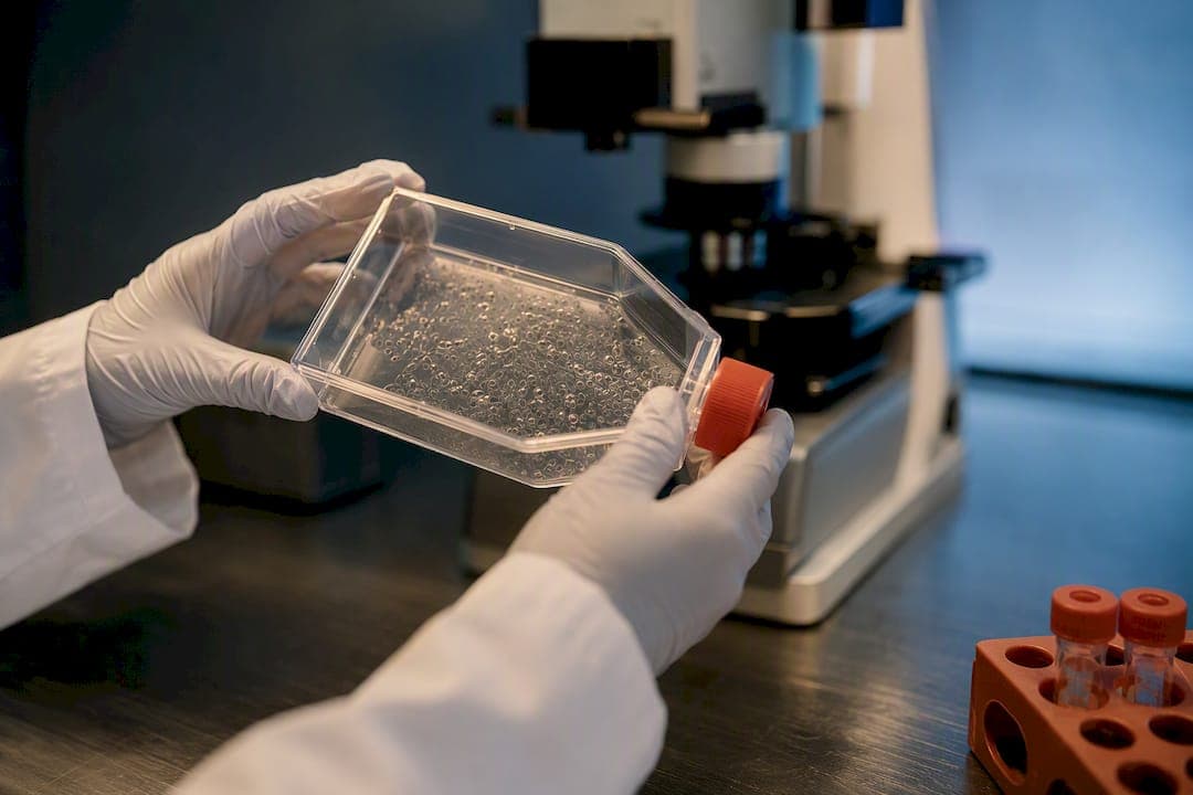
For most labs, the default choice is a two-step cytokine protocol: differentiate hematopoietic progenitor cells (HPCs) from iPSCs, then mature them with M-CSF, IL-34, and TGFβ-1 over roughly four to six weeks. If speed matters more than lineage fidelity, transcription-factor (TF) driven induction can deliver usable microglia-like cells in about two weeks. Either route, confirm identity with P2RY12 and TMEM119, not IBA1 alone, before trusting your data.
TL;DR:
- Cytokine protocols provide highly transcriptomic-matching microglia but require four to six weeks, with significant hands-on culture time and multiple harvests.
- Transcription-factor-driven methods produce functional microglia in about two weeks but may introduce artifacts from genetic modification and require BSL2 infrastructure.
- Simplified, xeno-free protocols reduce complexity and cost, matching cytokine protocols’ timeframes, and are ideal for translational applications.
- Confirm microglial identity using P2RY12 and TMEM119 markers alongside functional assays, as IBA1 alone is insufficient for validation.
- Batch variability and marker discordance are common failure points, making replication and proper QC crucial for reliable microglia generation.
Table of Contents
- What Method Should You Use to Generate Microglia From iPSC Lines?
- Cytokine vs. TF-Driven vs. Xeno-Free: Which Protocol Wins?
- A Day-by-Day Checklist for Cytokine-Based Microglia Differentiation
- Which Markers Prove You Actually Made Microglia?
- How Do Labs Use iPSC-Derived Microglia in Practice?
- Why Did My Microglia Differentiation Fail?
- How RareLabs Applies These Protocols to Rare Disease Research
- Where This Field Is Actually Headed
- Get Patient-Derived Microglia Models Without Building the Pipeline Yourself
- Where to Find the Full Protocol Details
- Sources
- FAQ
What Method Should You Use to Generate Microglia From iPSC Lines?
Microglia are not derived from neuroectoderm like neurons or astrocytes. They originate from yolk-sac primitive hematopoiesis during embryonic development, migrating into the brain before the blood brain barrier closes. That single fact explains why every credible protocol for generating microglia from iPSCs routes through a hematopoietic intermediate rather than a neural one. Skip that step, and you get macrophage-like cells with the wrong developmental address.
Three protocol families have emerged from this shared starting point, and each answers a different experimental question.
- Cytokine-driven two-step protocols mimic developmental biology closely, pushing iPSCs through primitive hematopoiesis into HPCs, then maturing those HPCs with defined cytokines. These produce cells with strong transcriptomic overlap to primary human microglia.
- Transcription-factor-driven rapid induction bypasses much of the slow cytokine choreography by overexpressing myeloid master regulators like PU.1/SPI1, forcing iPSCs toward a microglial fate in a fraction of the time.
- Simplified or xeno-free protocols, sometimes called second-generation or "2.0" methods, strip out complex requirements like hypoxia chambers or fluorescence-activated cell sorting (FACS) while keeping the cells transcriptionally comparable to first-generation methods.
Choosing among them comes down to your endpoint. Basic biology and mechanistic studies benefit from the developmental fidelity of cytokine-based differentiation. High-throughput screening favors TF-driven speed, since running a full six-week protocol across a 384-well drug library is rarely practical. Translational or clinical-adjacent work leans toward xeno-free adaptations, because animal-derived serum components complicate any path toward IND-enabling data packages. None of these approaches is universally correct. The right one depends on whether you are answering a biological question, screening compounds, or building toward a therapeutic program.
Cytokine vs. TF-Driven vs. Xeno-Free: Which Protocol Wins?
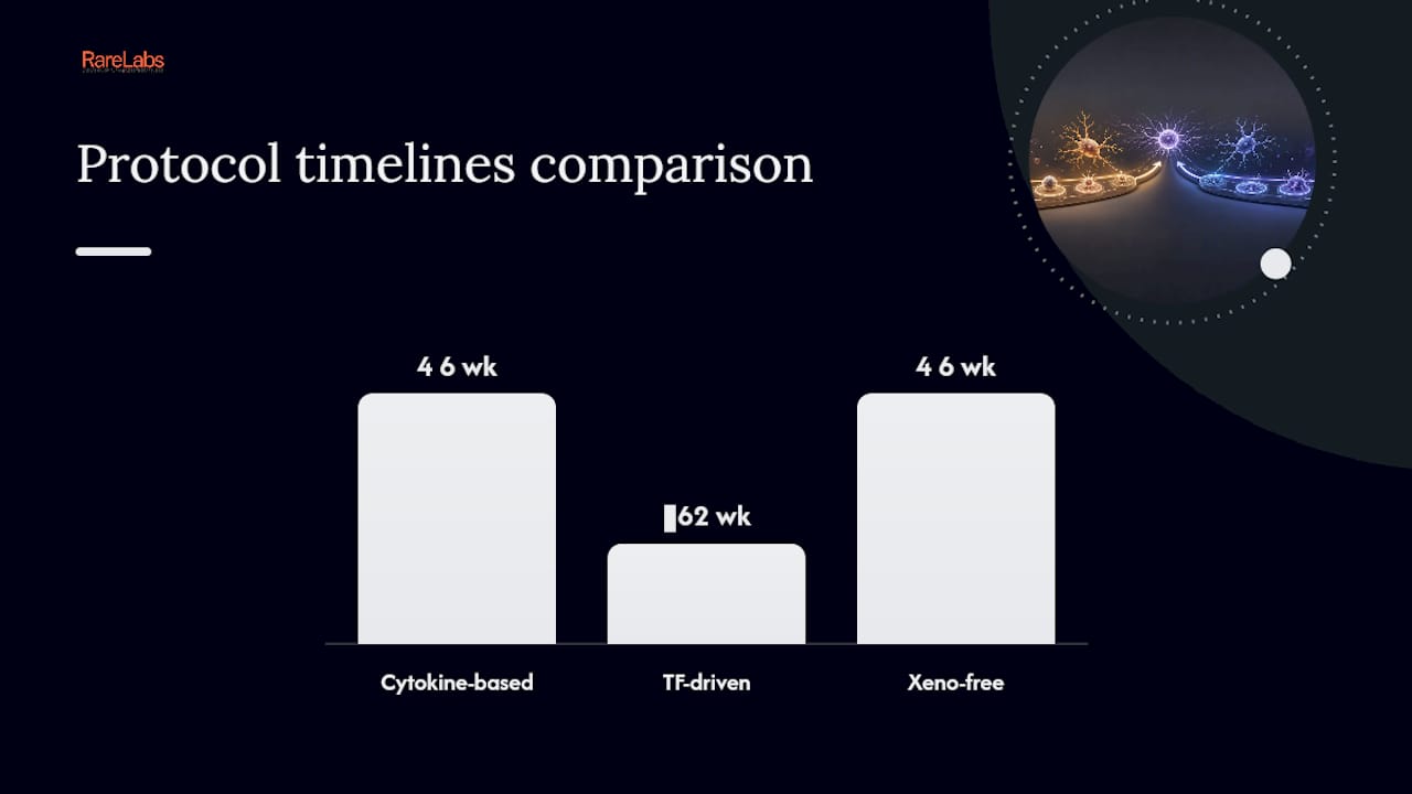
Timeline is usually the first filter labs apply, and it separates these three families cleanly. Cytokine-based two-step protocols typically run four to six weeks from iPSC to mature microglia, sometimes longer depending on line-specific kinetics. TF-driven methods compress that timeline to about two weeks by forcing myeloid identity directly rather than waiting for cells to pass through natural hematopoietic checkpoints. Xeno-free simplified protocols generally require a time comparable to cytokine-based methods but with reduced hands-on complexity.
Cytokine-based two-step differentiation remains the most cited and most validated approach in the literature. The Abud et al. protocol established the template still used across dozens of labs: induce HPCs from iPSCs using cytokines that mimic primitive hematopoiesis, then transition those floating progenitors into adherent, ramified microglia using a maturation cocktail built around M-CSF, IL-34, and TGFβ-1. The resulting cells express core microglial identity genes and respond to canonical stimuli in functional assays. Reagent costs run moderate, since M-CSF and IL-34 are standard recombinant cytokines available from most suppliers, and no genetic engineering step is required. The tradeoff is time and hands-on labor. Expect multiple media changes per week and a maturation window that demands patience.
TF-driven rapid induction answers a different question: how fast can you get functional cells at scale? A single transcription factor approach published in Inflammation and Regeneration demonstrated that overexpressing a myeloid master regulator can push iPSCs to functional microglia far faster than waiting on natural developmental cues. A newer xeno-free protocol built on the KOLF2.1J reference line pushes this further, generating usable microglia in as little as two weeks using inducible transcription factor strategies. The catch is infrastructure. These systems require stable integration or transduction of inducible constructs, which means BSL2 genetic-engineering practices, antibiotic selection steps, and extra quality control to rule out transgene artifacts on baseline inflammatory tone. If your lab already runs CRISPR or lentiviral work routinely, this cost is manageable. If it doesn't, the two-step cytokine method is the lower-friction entry point.
Simplified and xeno-free adaptations target a specific pain point: dependence on hypoxia chambers, FACS sorting, or co-culture setups that many labs simply don't have. The iPS-microglia 2.0 method validated that removing these requirements doesn't sacrifice transcriptional similarity to earlier, more complex protocols. One practical detail worth knowing: the small molecule IDE1 can substitute for recombinant TGFβ-1 in some conditions, cutting reagent cost and lot-to-lot variability that comes with recombinant growth factors. For labs building toward translational work, avoiding animal-derived serum components early also simplifies any future conversation about IND-enabling documentation.
| Protocol family | Typical timeline | Infrastructure needs | Best fit |
|---|---|---|---|
| Cytokine two-step | ~4–6+ weeks | Standard tissue culture, no genetic engineering | Basic biology, functional fidelity |
| TF-driven rapid | ~2 weeks | BSL2, transduction/integration capability | High-throughput screening |
| Xeno-free simplified | ~4–6 weeks, fewer steps | Standard culture, no hypoxia/FACS required | Translational, resource-limited labs |
None of these is a drop-in replacement for the others. A lab running a mechanistic neuroinflammation study should not default to TF-driven speed just because it's faster, since the forced overexpression can leave transgene-related artifacts sitting in the background of your inflammatory readouts. Conversely, a screening core testing 3,000 compounds against a single genetic target has no business running six-week cytokine protocols for every plate.
A Day-by-Day Checklist for Cytokine-Based Microglia Differentiation
Running the two-step cytokine protocol without a clear checkpoint map is how most labs lose weeks to unexplained variability. Here's a compact version built from the Abud et al. framework and practical optimizations reported in a recent review of microglia differentiation methods.
Materials to have on hand before starting:
- iPSC line with confirmed pluripotency and normal karyotype
- Hematopoietic induction medium (commercial kit or custom formulation)
- M-CSF, IL-34, and TGFβ-1 (or IDE1 as a substitute) for maturation
- Laminin-521 or poly-D-lysine plus laminin-521 coated plates
- Low-attachment plates for the HPC floating stage
- Weeks 0 to 2: Seed iPSCs and begin hematopoietic induction. Monitor for the emergence of floating, round HPCs, which typically start appearing toward the end of week two.
- Weeks 2 to 4: Harvest floating HPCs sequentially rather than in one pass. Collecting supernatant every 2 to 3 days over multiple harvest windows meaningfully increases total HPC yield compared to a single collection.
- HPC transfer: Plate harvested HPCs onto laminin-coated surfaces (or uncoated glass, per some optimizations) in maturation medium containing M-CSF, IL-34, and TGFβ-1.
- Weeks 4 to 6: Watch for morphological transition. Cells should flatten, extend processes, and adopt the ramified appearance associated with resting-state microglia.
- First QC checkpoint (around week 5 to 6): Run flow cytometry or immunocytochemistry for P2RY12 and TMEM119 before committing cells to downstream assays.
- Functional validation window: Cells are typically usable for functional assays anywhere from 7 to 60 days post-maturation, depending on the specific readout you need.
- Cryopreservation decision point: If you're not using cells immediately, freeze at the HPC stage rather than after full maturation. HPCs bank and recover more reliably than fully differentiated, adherent microglia.
Pro Tip: Bank HPCs from at least two to three independent differentiation runs per iPSC line before starting a large experiment. Line-to-line and batch-to-batch variability in hematopoietic yield is one of the most underestimated failure points in this entire workflow, and having backup vials saves you from restarting a six-week protocol from scratch.
Plastic adhesion losses are a quieter problem worth flagging. Microglia-like cells can stick tenaciously to certain plasticware during media changes, and aggressive pipetting at the wrong stage strips out a meaningful fraction of your culture before you ever reach the assay stage. Gentle, wide-bore pipetting during early maturation stages preserves more cells for downstream work.
Which Markers Prove You Actually Made Microglia?
TMEM119 and P2RY12 are the two markers that separate a genuine microglia identity claim from a maybe. Neither is optional, and neither should be used in isolation.
Protein and gene markers to run:
- P2RY12 and TMEM119: homeostatic markers specific to microglia, not shared broadly with peripheral macrophages
- IBA1: a pan-myeloid marker, useful for general identification but not specific to microglia versus other tissue macrophages
- PU.1 (SPI1): master transcription factor confirming myeloid lineage commitment
- TREM2 and HEXB: functional and homeostatic markers increasingly used in microglia signature panels
The caveat that trips up newer labs: IBA1 and CD68 positivity alone does not confirm microglial identity. Both markers are shared broadly across macrophage populations, so a culture that's IBA1-positive but TMEM119-negative is more honestly described as macrophage-like, not microglia. This distinction matters most in papers making disease-relevance claims, where reviewers increasingly expect the homeostatic marker panel alongside pan-myeloid staining.
Functional assays close the gap that markers alone leave open. A protocol validated for phagocytic function confirmed that properly differentiated cells take up pHrodo-labeled bioparticles and respond to canonical stimuli like LPS with appropriate cytokine release. Beta-amyloid uptake assays extend this further for labs working in neurodegeneration contexts, since impaired amyloid clearance is a core hypothesis in Alzheimer's disease research involving microglia function.
Transcriptomic benchmarking rounds out a rigorous validation package. Running PCA or signature-score comparisons against published fetal and adult human microglia RNA-seq datasets tells you how closely your cells track the real biology, beyond what any five-marker panel can capture. Labs publishing in higher-tier journals increasingly include this comparison as a baseline expectation, not a nice-to-have.
How Do Labs Use iPSC-Derived Microglia in Practice?
Neuroinflammation modeling, target validation, and drug screening account for most current applications of iPSC-derived microglia in disease research. But the experimental design details shift substantially depending on whether you're running a co-culture, a screen, or an in vivo transplant.
Co-culture design is where most synapse pruning and neuroinflammation studies live. Timing the introduction of microglia relative to neuronal maturation matters enormously. Introduce microglia too early into an immature neuronal culture and you risk confounding developmental effects with genuine pruning behavior. Media compromises are unavoidable here: neurons and microglia have different optimal formulations, so most labs settle on a hybrid medium that keeps both populations viable, even if neither is at its individual optimum. Readouts typically center on synaptic marker colocalization (PSD95, synaptophysin) inside microglial processes, or cytokine release panels when modeling neuroinflammatory states.
Screening design is where the TF-driven versus cytokine-based decision has the most practical weight. A screen testing hundreds or thousands of compounds against a single genetic target benefits from the compressed timeline of TF-driven differentiation, since running that volume through a six-week cytokine protocol multiple times isn't realistic for most budgets. The tradeoff is that TF-driven cells need extra baseline QC to confirm the transgene itself isn't skewing your inflammatory readout before you ever add a compound.
- Match your differentiation method to your assay window, not the other way around.
- For long assay windows (weeks of live imaging), cytokine-based cells with a proven functional lifespan hold up better.
- For single-timepoint endpoint assays across large compound libraries, TF-driven speed usually wins.
Transplantation and in vivo integration studies push microglia into mouse models, often immunodeficient or humanized strains, to study long-term integration and function in a living brain environment. Endpoints typically include engraftment percentage, morphological ramification in situ, and response to induced injury or amyloid pathology. This is the highest-bar application, and it generally demands the developmental fidelity of cytokine-based or xeno-free protocols rather than TF-driven shortcuts, since transgene artifacts become harder to control for over months of in vivo residence.
Why Did My Microglia Differentiation Fail?
Most failed differentiations trace back to one of three root causes, and none of them require starting over from iPSCs if you catch them early enough.
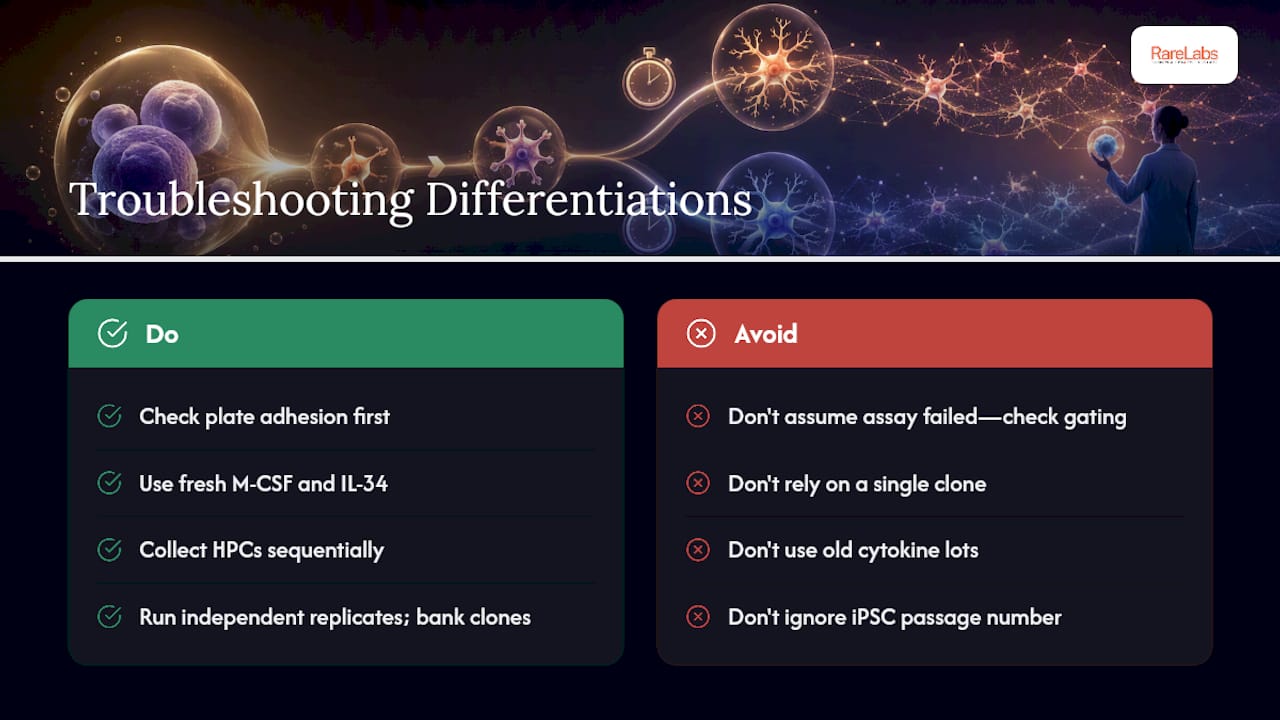
Low yield or unexpected HPC loss. Check plate adhesion first. If your floating HPC counts drop sharply between induction days, cells may be adhering to culture plastic rather than staying in suspension where they belong at this stage. Reagent freshness is the second suspect. M-CSF and IL-34 lose activity faster than labs often expect, and a subtly degraded cytokine lot produces a slow, frustrating decline in yield rather than an obvious failure. Sequential harvesting, collecting HPCs across multiple windows instead of one bulk collection, consistently improves total yield and is worth building into your standard workflow from the start.
Marker discordance. If your cells look ramified and express IBA1 but come back negative or dim for TMEM119 and P2RY12, don't assume the assay failed before checking your gating strategy and antibody clone. Some commercial antibody clones perform inconsistently across fixation protocols. When results stay ambiguous, transcriptional benchmarking against reference microglia datasets gives a more definitive answer than protein staining alone.
Line-to-line variability. This is the quiet source of irreproducibility that undermines otherwise solid experiments. Best practice is running biological replicates from independent differentiation batches, incorporating isogenic controls wherever possible (particularly important in disease modeling work), and banking multiple clones per patient line rather than relying on a single derivation.
Pro Tip: Before troubleshooting a "failed" differentiation, check the passage number of your starting iPSCs. Highly passaged lines drift genetically and functionally in ways that specifically compromise hematopoietic potential, often well before other differentiation lineages show any problem.
How RareLabs Applies These Protocols to Rare Disease Research
Running these protocols reliably at scale is a different challenge than running them once for a methods paper. A specialized biotechnology lab builds patient-derived iPSC disease models for ultra-rare and undiagnosed genetic conditions as part of a broader pipeline that includes CRISPR-edited isogenic controls, high-throughput screening of FDA-approved drugs, and custom antisense oligonucleotide development for mechanistic rescue testing.
For a rare neurodegenerative or neuroinflammatory condition, that pipeline typically starts with patient-derived iPSC banking, moves through targeted differentiation—including microglia where the disease biology calls for it—and feeds into parallel treatment screening designed to surface actionable therapeutic candidates faster than a single-target academic study usually can. Proprietary RINAE AI-based scoring supports target amenability assessment alongside the wet-lab screening, helping prioritize which mutations and pathways are worth pursuing first.
Foundations, physician-scientists, and biopharma partners working on microglia-relevant rare diseases can treat this as a direct service option rather than a purely internal capability to replicate. The role of pluripotent stem cells in translational disease modeling illustrates how patient-specific models feed directly into actionable treatment discovery.
Where This Field Is Actually Headed
The direction of travel in microglia differentiation isn't subtle: everything is moving toward faster, cleaner, more clinically compatible methods. TF-driven acceleration will keep closing the gap with cytokine-based fidelity as researchers refine which combinations of transcription factors minimize transgene artifacts. Xeno-free adaptations will keep gaining ground for one practical reason: nobody wants to explain animal-derived serum components to a regulatory reviewer if they can avoid it. Organoid and 3D co-culture integration is the next real frontier, since flat 2D cultures still miss the spatial and cellular context that shapes real microglial behavior in a living brain.
If you're choosing a path today, here's the honest breakdown: a small academic lab asking a mechanistic question should default to the cytokine two-step method for its fidelity and accessibility. A translational program building toward IND-enabling data should look hard at xeno-free variants from day one, not retrofit them later. A screening core running high volume should invest in TF-driven infrastructure once, then reap the speed advantage across every subsequent project.
What gets underestimated across all three paths is validation discipline. Teams that skip the marker and functional assay checkpoints tend to publish results that don't replicate, and the field has enough of those already.
— John
Get Patient-Derived Microglia Models Without Building the Pipeline Yourself
Building a validated microglia differentiation pipeline from scratch means months of protocol optimization before you ever touch your actual research question. Hopeatrarelabs skips that build phase entirely: patient-derived iPSC models, CRISPR-edited isogenic controls, and high-throughput drug screening already exist as a working pipeline, so a rare disease program can move straight to generating usable data instead of troubleshooting HPC yields for a quarter.

That matters most for foundations and families racing against disease progression, and for biopharma teams that need translational-quality models without staffing an entire stem cell core. The lab offers patient-derived iPSC disease model development and biobanking, high-throughput drug screening, custom ASO design and in vitro rescue validation, and IND-enabling data package preparation, all built around rigorous scientific standards. If your rare disease program needs a microglia model, or any other patient-specific cellular model, as part of a treatment discovery effort, visit the RareLabs services page to start an Amenability Study and Discovery Roadmap tailored to your specific mutation or condition.
Where to Find the Full Protocol Details
For complete reagent lists and step-by-step parameters, consult the primary literature directly rather than relying on summaries alone.
- iPSC-derived human microglia-like cells to study neurological diseases (Abud et al.)
- Efficient derivation of microglia-like cells from human pluripotent stem cells
- Simplified method to generate human microglia (iPS-microglia 2.0)
- Microglia differentiation protocol entry, protocols.io
Sources
- A xeno-free protocol for rapid differentiation of human iPSC-derived microglia from the KOLF2.1J reference line (preprint / protocol description)
- iPSC-derived human microglia-like cells to study neurological diseases (Abud et al., 2017)
- Efficient derivation of microglia-like cells from human pluripotent stem cells (protocol paper)
- Development and validation of a simplified method to generate human microglia from pluripotent stem cells (iPS-microglia 2.0)
FAQ
What Are the Downsides of Using iPSCs for Microglia Models?
iPSC-derived microglia can show significant line-to-line and batch-to-batch variability, and cells derived through TF-driven methods carry a risk of transgene-related artifacts affecting baseline inflammatory signaling. Marker discordance is also common if labs rely on pan-myeloid markers like IBA1 without confirming homeostatic identity through P2RY12 and TMEM119.
What Is the iPSC-Derived Microglia Protocol Used For?
These protocols generate patient-specific or disease-relevant microglia for modeling neuroinflammation, testing drug candidates, and studying microglia function in neurodegeneration without relying on scarce primary human tissue. This approach is applied as part of broader patient-derived disease modeling for rare neurological conditions.
Can Microglia Regenerate?
Yes, microglia can self-renew and repopulate the brain after depletion, largely through local proliferation of surviving cells rather than significant replacement from peripheral blood monocytes. This regenerative capacity is part of why researchers study microglial turnover dynamics using iPSC-derived models alongside in vivo systems.
What Are the Two Main Types of Microglia Discussed in Differentiation Work?
Researchers typically distinguish homeostatic (resting-state) microglia, marked by P2RY12 and TMEM119 expression, from activated or disease-associated microglia states that upregulate inflammatory and phagocytic gene programs. Differentiation protocols aim to produce the homeostatic phenotype first, then use stimuli like LPS or beta-amyloid to model the activated state.
How Long Does It Take to Generate Microglia From iPSC Lines?
Cytokine-based two-step protocols typically take four to six weeks or longer, while transcription-factor-driven rapid methods can produce usable microglia in about two weeks. The right timeline depends on whether your experiment prioritizes developmental fidelity or throughput speed.