
For fast, high-purity glutamatergic neurons, use inducible NGN2 transcription-factor induction: expect functional neurons in roughly 14 days. For region-specific or developmentally staged neurons, dual-SMAD inhibition is the better route, with neural rosettes forming around days 12 to 15 and terminal maturation stretching past six weeks. Skip NGN2 when your experiment needs subtype diversity or organoid-level patterning; skip dual-SMAD when your screen needs speed and homogeneity.
TL;DR:
- NGN2 protocols produce glutamatergic neurons within 14 days with high purity, ideal for screening and rapid validation.
- Dual-SMAD inhibition yields more diverse and developmentally accurate neuron subtypes but takes 4 to 6 weeks to reach maturity.
- Clone selection and genomic validation, especially using array CGH, are critical to avoid inconsistent differentiation outcomes.
- Neuronal functional maturity, such as synapse formation and electrophysiological activity, typically occurs between 6 to 9 weeks in culture.
- For patient-specific modeling, standardization of workflow and endpoints ensures reproducibility across batches and studies.
Table of Contents
- What Is Neuronal Differentiation From iPSCs?
- How Do You Run an NGN2 Transcription-Factor Protocol?
- What Does a Dual-SMAD Rosette Protocol Look Like?
- How Long Does It Take Neurons to Mature Functionally?
- What Causes Failed or Inconsistent Neuronal Differentiation?
- How Does RareLabs Standardize Neuronal Differentiation for Patient Models?
- Which Protocol Should You Actually Run First?
- How Can Hopeatrarelabs Support Your Neuronal Modeling Project?
- Sources
What Is Neuronal Differentiation From iPSCs?
Neuronal differentiation from induced pluripotent stem cells is the process of converting patient-derived or reference iPSC lines into post-mitotic neurons using either forced transcription-factor expression or staged small-molecule signaling. Both routes exist because they solve different problems, and the choice you make on day zero shapes everything downstream, from how many replicates you can run to whether your neurons resemble anything found in an actual developing brain.
The two dominant frameworks are TF-driven induction, most commonly built on doxycycline-inducible NGN2, and dual-SMAD inhibition, which blocks BMP and TGFβ signaling to mimic early neural tube development. Neither is objectively "better." One optimizes for speed and purity, the other for biological fidelity and subtype range. Here's how they stack up on the axes that actually matter at the bench.
Efficiency and purity. NGN2 protocols routinely report near-complete conversion to glutamatergic neurons within a two-week timeline, with minimal residual progenitor contamination. Dual-SMAD cultures are messier at the intermediate stage. Rosette-derived NPC populations carry more heterogeneity, and purity depends heavily on how aggressively you enrich for PAX6+/NESTIN+ cells before expansion.
Timeline. NGN2 gets you to a countable, stainable neuron in about two weeks. Dual-SMAD needs 12 to 15 days just to reach rosette formation, then several more weeks of patterning and expansion before you have anything resembling a mature post-mitotic neuron.
Subtype scope. This is where dual-SMAD wins outright. Add sonic hedgehog agonists or retinoic acid to a dual-SMAD backbone and you can push toward dopaminergic, motor neuron, or GABAergic fates. NGN2 alone gives you cortical glutamatergic neurons, full stop, unless you layer in additional transcription factors like ASCL1 or ISL1/LHX3 for motor neuron identity.
Scalability. NGN2's short timeline and single induction step make it far more automation-friendly for screening pipelines. Dual-SMAD's multi-week, multi-media protocol is harder to standardize across plates and operators.
- NGN2: fast, pure, screening-ready, limited subtype range
- Dual-SMAD: slower, more variable, but developmentally faithful and subtype-flexible
Pro Tip: If your assay depends on comparing isogenic lines at scale, run NGN2 first to validate your hit list, then confirm top candidates in dual-SMAD-derived subtype-specific neurons before committing to animal studies.
How Do You Run an NGN2 Transcription-Factor Protocol?
The NGN2 workflow is a single, tightly scheduled induction rather than a staged developmental recapitulation. Here's the practical sequence most labs run, adapted from rapid single-step induction protocols.
Before you start, confirm your iPSC line carries a doxycycline-inducible NGN2 cassette, either through lentiviral transduction or, preferably, safe-harbor integration at a locus like AAVS1. You'll also want puromycin or a fluorescent reporter for selection, laminin-coated plates, and a defined neuronal maturation medium (typically Neurobasal plus B27 and BDNF/GDNF/NT3).
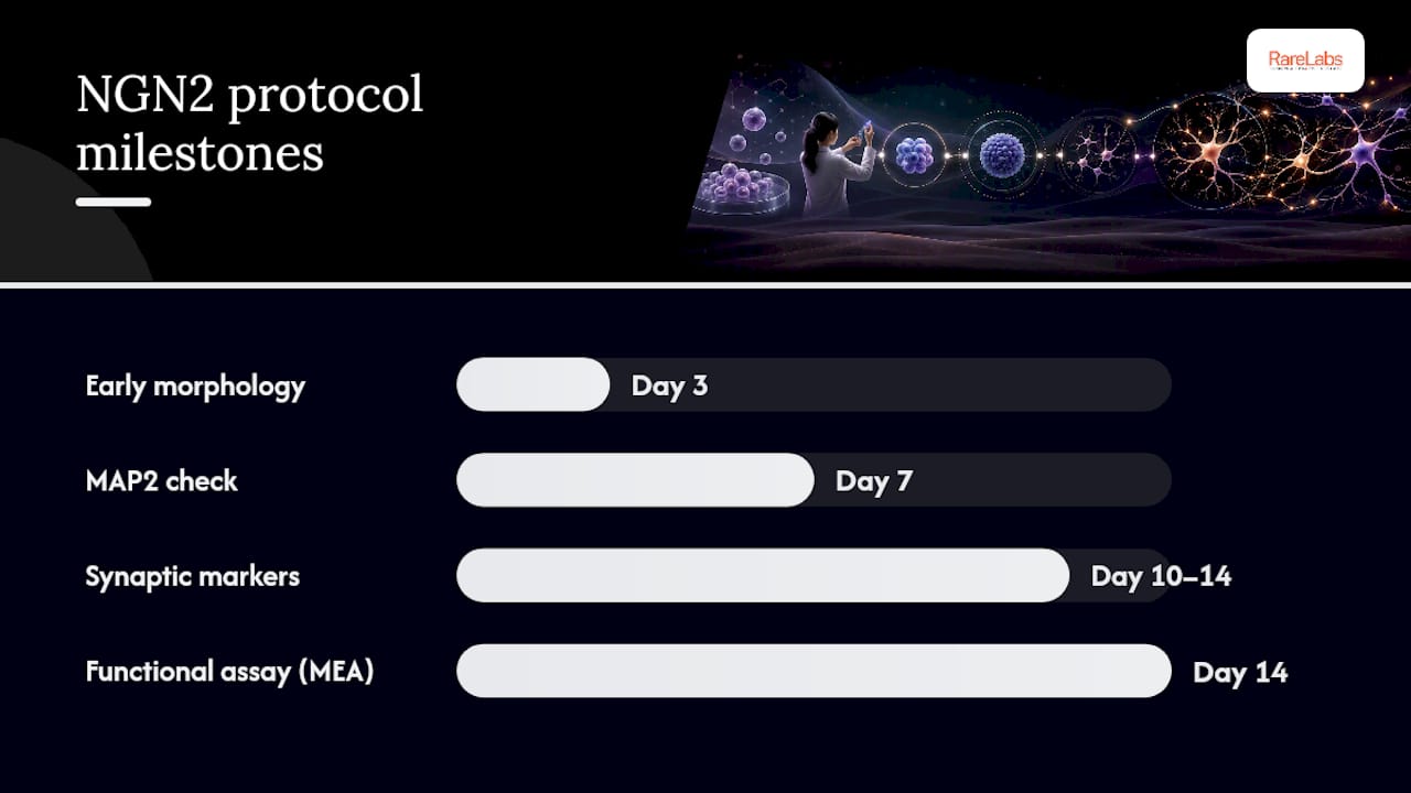
- Day 0: Dissociate iPSCs to single cells and plate at high density on laminin-coated dishes in induction medium containing doxycycline (typically 2 µg/mL) to activate NGN2 expression.
- Day 1 to 2: Add puromycin selection if your construct includes a resistance cassette, eliminating non-transduced cells and enriching for uniform NGN2 expression.
- Day 3: Confirm morphology shift toward bipolar, neurite-bearing cells. This is a good checkpoint for early NESTIN downregulation and initial βIII-Tubulin expression by immunostaining.
- Day 4 to 7: Switch to neuronal maturation medium, often co-culturing with mouse glia or human astrocytes to support synaptic development. Reduce doxycycline gradually or maintain depending on your construct's design.
- Day 7: Check MAP2 and βIII-Tubulin expression. You should see extensive neurite outgrowth and largely resolved proliferative markers.
- Day 10 to 14: Full media changes every 2 to 3 days with maturation supplements. Cells should show Synapsin puncta and early PSD-95 expression by immunostaining.
- Day 14: Evaluation of MAP2+ purity and functional activity by multi-electrode array (MEA) recordings can be conducted at this stage.
Statistic Callout: Standard NGN2 protocols achieve differentiation efficiency and purity approaching 100% in many published reports within a two-week window, a number dual-SMAD protocols rarely match at the same timepoint.
Delivery method matters more than most people assume. Safe-harbor, site-specific integration produces more uniform TF expression across clones than random lentiviral integration, which translates directly into tighter data in dose-response screens. mRNA-based delivery avoids genomic integration entirely and suits one-off experiments, but it sacrifices the reproducibility of a stable line if you plan to run the same differentiation dozens of times across a project.
Pro Tip: Quantify NGN2 expression variance across your candidate clones by qPCR before scaling up. A clone with tight expression variance will save you weeks of troubleshooting inconsistent maturation later.
What Does a Dual-SMAD Rosette Protocol Look Like?
Dual-SMAD inhibition earns its complexity by tracking actual neural tube development, which is exactly why it takes longer and produces more diverse cell types.
The core chemistry blocks two signaling arms simultaneously: SB431542 inhibits TGFβ/Activin signaling, while Noggin or its small-molecule substitute LDN193189 blocks BMP signaling. Together they push iPSCs toward a default neuroectodermal fate instead of mesoderm or endoderm.
- Day 0 to 1: Plate iPSCs as a monolayer or in embryoid bodies, then begin dual-SMAD inhibition with SB431542 (10 µM) and LDN193189 (100 nM) in a neural induction medium.
- Day 4 to 6: Continue daily media changes with both inhibitors. PAX6 expression should begin rising as pluripotency markers OCT4 and NANOG decline.
- Day 8 to 11: Neuroepithelial sheets become visible. Some protocols use manual "spot plating," seeding cells in discrete colonies to encourage synchronized rosette formation rather than a continuous sheet.
- Day 12 to 15: Neural rosettes form, visible as radial, tube-like structures under phase microscopy, a consistent developmental milestone across dual-SMAD protocols. Confirm PAX6+/NESTIN+ identity by immunostaining.
- Day 15 to 20: Manually or enzymatically pick rosettes, then dissociate with Accutase for NPC expansion. Add mitotic inhibitors sparingly if overproliferation crowds out differentiation.
- Week 3 to 4: Expand NPCs in FGF2/EGF-supplemented medium. This is the ideal window to cryobank a large NPC stock for synchronized future experiments.
- Week 4 onward: Withdraw growth factors and switch to neuronal differentiation medium, optionally adding subtype-specifying morphogens (sonic hedgehog for motor neurons or ventral fates, Wnt agonists for dorsal identities).
- Week 6 to 10: Terminal neuronal markers (MAP2, βIII-Tubulin, subtype-specific transcription factors) should be robust, though full functional maturity often takes longer.
Plating density matters more here than in NGN2 work. Overcrowded rosettes fuse and lose radial organization; underseeded plates yield patchy, inconsistent neuroepithelium. Most labs settle on 40,000 to 80,000 cells/cm² at induction, adjusted per line.
- Cryobank NPCs at the day 20 to 25 window once PAX6/NESTIN identity is confirmed
- Refresh media every 2 days during induction, every 3 to 4 days during expansion
- Re-check pluripotency markers (OCT4, NANOG) at day 6 to confirm exit from the stem state
How Long Does It Take Neurons to Mature Functionally?
Morphology arrives faster than function, and that gap trips up a lot of otherwise well-run experiments. Neurons can look mature under a microscope, with extensive MAP2-positive neurites, weeks before they fire reliable action potentials or form functional synapses.
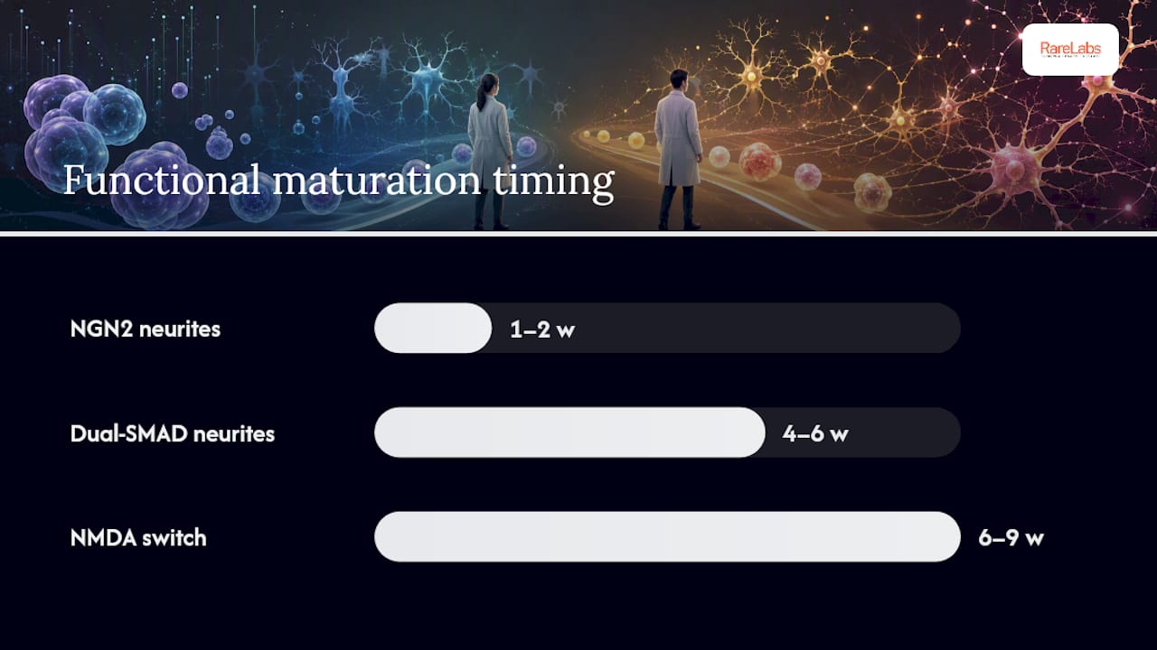
The general sequence runs: neurite outgrowth and MAP2/βIII-Tubulin expression first (week 1 to 2 for NGN2, week 4 to 6 for dual-SMAD), followed by Synapsin and PSD-95 punctate expression signaling synapse formation, then finally coordinated electrical activity. Synaptic maturation and NMDA receptor subunit switching typically appear after several weeks in culture, commonly observed between six and nine weeks, regardless of which induction method you started with.
Statistic Callout: TF-induced neurons in optimized conditions can show dendritic spine formation and NMDA subunit switching by roughly day 60 to 75, putting NGN2-derived cultures on a faster track to postnatal-like maturity than most organoid systems.
Three assay types tell you whether your neurons are ready for downstream work:
- Immunocytochemistry for Synapsin-1 and PSD-95 confirms structural synapse formation but says nothing about function.
- Multi-electrode array (MEA) recording captures network-level spontaneous activity and burst synchrony, ideal for screening throughput.
- Patch clamp electrophysiology gives the gold-standard readout, resting membrane potential, action potential firing, and evoked synaptic currents, but it's low-throughput and technically demanding.
A negative MEA result at week 4 isn't necessarily a failure. It often just means you're too early. A negative result at week 9, paired with weak Synapsin staining, usually means the culture needs intervention: astrocyte co-culture, BDNF/GDNF supplementation, or a longer maturation window. Reporting molecular markers and electrophysiology together avoids the false sense of maturity that marker panels alone can create.
What Causes Failed or Inconsistent Neuronal Differentiation?
Most differentiation failures trace back to one of three sources: the starting iPSC line, inconsistent TF expression, or undetected genomic damage. Fixing the right one saves weeks.
Genomic integrity comes first. Standard karyotyping may miss sub-chromosomal rearrangements that can affect differentiation efficiency that can silently impair differentiation efficiency weeks into a protocol. Run array comparative genomic hybridization or shallow whole-genome sequencing on any line before committing it to a long dual-SMAD timeline.
Line-to-line and clone-to-clone heterogeneity shows up as inconsistent rosette formation or patchy NGN2 induction. For TF lines, qPCR-quantify inducible expression across candidate clones early and retire outliers before scaling.
- Poor survival at induction: check seeding density and laminin coating quality first
- Incomplete differentiation at day 14 (NGN2): verify doxycycline concentration and selection stringency
- Failed rosette formation (dual-SMAD): re-check SB431542/LDN193189 potency and lot consistency
- Batch drift across experiments: cryobank NPCs at a validated stage to synchronize future differentiation starts
Pro Tip: Keep a frozen NPC bank from a single, genomically validated passage. Thawing from one synchronized stock removes an entire category of variability from multi-month screening campaigns.
How Does RareLabs Standardize Neuronal Differentiation for Patient Models?
Turning a working protocol into a reproducible screening platform means building checkpoints into the workflow itself, not just following the steps once and hoping.
Hopeatrarelabs structures patient-derived neuronal modeling around a sequence built for repeatability: genomic QC first, then induction method selection matched to the disease question, then NPC banking for synchronized starts, then parallelized differentiation runs against standardized endpoint metrics. This mirrors what happens in drug candidate prioritization, where consistent endpoints across dozens of parallel wells matter more than any single elegant protocol.
- Genomic screening before committing a patient line to a multi-week protocol
- Method selection based on the disease's cell type of interest, not convenience
- NPC cryobanking to enable multi-site or longitudinal comparisons from an identical starting population
- Standardized endpoint metrics, such as percent MAP2+ at day 14 and MEA baseline activity, tracked across every run for cross-batch comparability
The goal isn't a single perfect experiment. It's a metric set stable enough that a result from March means the same thing as a result from October.
Which Protocol Should You Actually Run First?
If your study needs fast, homogeneous glutamatergic neurons for a compound screen or an isogenic comparison, run NGN2 first. It's faster, cheaper per experiment, and the safe-harbor integration approach gives you clone-to-clone consistency that dual-SMAD protocols struggle to match.
Reach for dual-SMAD when the biology itself demands it: motor neuron disease models, dopaminergic degeneration studies, or anything where regional identity is the whole point of the experiment. Budget your resources accordingly. A pilot screen doesn't need a six-week timeline; a mechanistic study of subtype-specific vulnerability usually can't skip it.
If you're weighing a patient-specific program against a general research question, that's worth a direct conversation before you commit months to either protocol.
— John
How Can Hopeatrarelabs Support Your Neuronal Modeling Project?
Running these protocols on a patient's own cells, at diagnostic speed, is a different operation than optimizing a reference line in a university lab. A specialized biotechnology lab builds patient-specific iPSC models and matches the differentiation method to the disease question rather than defaulting to whichever protocol is fastest to set up.

A typical engagement starts with banking two to three validated clones from the patient's own cells, followed by genomic screening, induction method selection (NGN2 for speed, dual-SMAD for region-specific fidelity), NPC banking for reproducible reruns, and parallelized screening against FDA-approved compounds, custom ASOs, or gene therapy candidates. Deliverables scale with the disease question, but every program is built around the same standardized endpoints described above, so results from patient-derived cells can be compared meaningfully against controls. If a family, foundation, or physician-scientist is weighing whether a personalized modeling program makes sense for a specific rare diagnosis, start a conversation with Hopeatrarelabs about what a program would look like for that case.