

Ex vivo drug testing exposes a patient's own living tissue, removed from the body but kept functionally intact, to drug candidates outside the body to measure how that specific tissue actually responds. The National Cancer Institute defines ex vivo as occurring "outside of the living body," typically on tissue, cells, or organs taken from a patient and sometimes returned afterward.
The method matters most in functional precision oncology and preclinical screening, where genomic data alone can't predict drug response. It fits best with patient-derived samples in refractory cancers, rare diseases, or undiagnosed conditions where clinical trial data barely exist. One caution up front: results are only as good as the tissue sample and the validation behind the assay.
- A 2026 review of 48 studies supports ex vivo models as a complement to genomic profiling.
- The SMARTrial hit its 7-day reporting endpoint in 91% of cases, with a 3-day median turnaround.
- Services like Hopeatrarelabs use this approach to screen thousands of drugs against patient-specific disease models.
Key Takeaways
Ex vivo drug testing works because it measures a patient's actual tissue response to treatment, capturing resistance and sensitivity signals that genomic sequencing alone cannot detect.
| Point | Details |
|---|---|
| Match model to disease | Organoids, slices, or perfused platforms each fit different research questions and disease types. |
| Turnaround varies by tissue | Hematologic samples average around 3 days; solid tumors can take 10 to 12 days. |
| Validate with prospective data | Look for independent cohort validation and documented repeatability before trusting a report. |
| Sample quality drives success | Cold ischemia time and viable tissue amount are the most common points of failure. |
| Personalized programs exist today | Hopeatrarelabs builds patient-specific disease models and runs parallel repurposing screens for rare diseases. |
Table of Contents
- What Is Ex Vivo Testing, and How Does It Differ From In Vitro and In Vivo?
- How Ex Vivo Drug Testing Works: Models and Workflow
- What Assays and Readouts Does Ex Vivo Testing Use?
- Where Ex Vivo Testing Adds the Most Translational Value
- How Reliable Is Ex Vivo Testing, and What Are Its Limits?
- Planning an Ex Vivo Study: Timelines, Costs, and Ethics
- A Patient-Specific Screening Program in Practice
- Where Ex Vivo Testing Fits Now
- How to Access a Patient-Specific Screening Program
- Frequently Asked Questions
- Sources
What Is Ex Vivo Testing, and How Does It Differ From In Vitro and In Vivo?
"Ex vivo" gets thrown around loosely, but the term carries a specific meaning: tissue removed from a living organism and studied in conditions that preserve its native architecture, cell-to-cell contacts, and, when possible, its microenvironment. That's a meaningful distinction from generic cell culture. Tissue engineering researchers have pushed back on treating ex vivo and in vitro as interchangeable, since ex vivo work is defined by its attempt to keep the tissue behaving as it would inside the body, not just alive in a dish.
Here's how the three approaches stack up:
- In vitro: Cells grown in plastic dishes, often as 2D monolayers or immortalized lines. High throughput, low cost, low physiological relevance.
- Ex vivo: Intact or minimally disrupted tissue from a living donor, tested outside the body. Preserves architecture and often immune or stromal components. Moderate throughput, higher fidelity.
- In vivo: Whole-organism testing, usually animal models or clinical trials. Highest physiological relevance, slowest and most expensive.
Choose ex vivo when the research question depends on tissue architecture, cell heterogeneity, or a patient-specific microenvironment that a cell line simply can't replicate. Genomic profiling can tell you what mutations exist. Ex vivo testing tells you what the tissue actually does when a drug hits it.
How Ex Vivo Drug Testing Works: Models and Workflow
Model choice drives everything downstream. Each format trades physiological fidelity against speed and sample requirements, and picking wrong is one of the most common reasons ex vivo programs stall.
Tissue slices and organotypic cultures preserve the most native architecture, keeping stromal cells, vasculature fragments, and immune infiltrate intact. They demand more tissue and skilled handling, and viability windows are short, but they generate the closest functional analog to the intact tumor or organ.
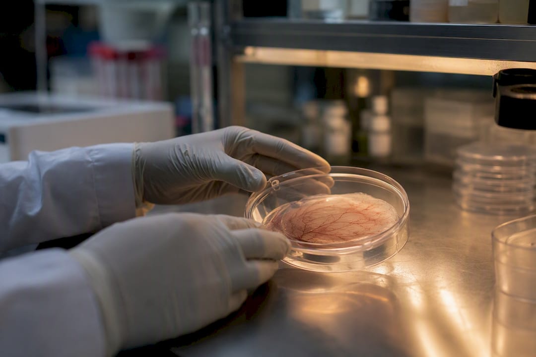
Organoids grow from patient-derived stem or progenitor cells into 3D structures that retain key histological and genetic features. Research on organoid fidelity shows they outperform 2D cultures for many tumor types, though they take longer to establish than slices.

Spheroids and multicellular clusters sacrifice some structural complexity for speed. They're faster to generate and easier to scale, which makes them useful when turnaround time outweighs the need for full microenvironment fidelity.
Dissociated primary cells offer the fastest setup and highest throughput but lose tissue architecture almost entirely, limiting insight into cell-cell signaling.
Perfused tissue platforms, exemplified by systems like Ex Vivo Metrics™, keep isolated tissue blood-perfused to sustain near-physiological function, useful for organ-level pharmacology questions where static culture falls short.
A standard workflow looks like this:
- Collect tissue via biopsy, surgical resection, or blood draw, minimizing cold ischemia time.
- Transport in validated media at controlled temperature, usually within hours of collection.
- Process the sample: dissociate, slice, or embed depending on model type.
- Establish culture and confirm baseline viability before any drug exposure.
- Expose tissue to drug panels across a dose range, often in parallel.
- Read out results using viability, imaging, or functional assays.
- Interpret data against reference ranges and correlate with clinical history where available.
Pro Tip: Don't treat transport media as an afterthought. A 2026 Nature Medicine analysis found that programs treating ex vivo models as "off-the-shelf," rather than matching media and handling protocols to the specific disease, account for a large share of operational failures.
What Assays and Readouts Does Ex Vivo Testing Use?
The readout you choose determines what question you can actually answer. Viability assays like ATP quantification or resazurin reduction measure bulk metabolic activity, and a sharp drop after drug exposure suggests cytotoxicity, though it won't tell you the mechanism. Apoptosis markers, such as caspase activation or Annexin V staining, narrow that down by confirming programmed cell death rather than necrosis.
High-content imaging, sometimes called pharmacoscopy, tracks individual cells over time and can reveal morphological changes invisible to bulk assays. Flow cytometry adds resolution across cell populations, useful when tissue contains a mix of tumor and immune cells and you need response broken out by cell type.
Single-cell transcriptomics goes further, capturing gene expression shifts that flag resistance mechanisms before they show up phenotypically. Functional assays like electrophysiology or contractility testing apply mainly to specialized tissue such as cardiac or neuronal samples, where the readout is a physiological function rather than survival.
- Combine at least two orthogonal readouts before drawing conclusions from a single assay.
- Layer omics data onto functional results when resistance patterns look ambiguous.
- Treat any single-assay result as preliminary until confirmed by a second method.
Where Ex Vivo Testing Adds the Most Translational Value
Ex vivo data earns its place in three parts of the pipeline: candidate prioritization, mechanism-of-action work, and combination screening. Instead of guessing which of ten drug candidates deserves further investment, a lab can expose patient-derived tissue to all ten in parallel and rank them by actual functional response. That's faster and often cheaper than running each candidate through a full in vivo study first.
Functional precision oncology takes this further by applying ex vivo screening directly to a patient's own tumor material to identify vulnerabilities that genomic sequencing misses entirely. A tumor might carry no actionable mutation on a genomic panel yet show striking sensitivity to a drug nobody would have predicted from DNA alone. Experts describe this functional readout as capturing drug-tumor interactions that genomic data simply can't see, a gap that matters most in rare or refractory cancers where trial evidence is thin.
The core value proposition of functional precision oncology isn't replacing genomics. It's catching the resistance and sensitivity signals that mutation calling alone leaves on the table, especially in tumors with no clear genomic driver.
Clinical-correlation studies typically follow one of two designs: prospective observational cohorts that compare ex vivo predictions against actual patient outcomes, or pilot-guided therapy studies where a tumor board actively uses the ex vivo report to select treatment. The SMARTrial falls into the first category, and its results showed that ex vivo drug resistance predicted treatment failure in hematologic malignancies with enough consistency to inform real clinical discussions.
- Candidate prioritization across drug libraries before committing to animal studies.
- Combination screening to spot synergy that single-agent testing would miss.
- Tumor board input where genomic testing alone left no actionable target.
How Reliable Is Ex Vivo Testing, and What Are Its Limits?
Ex vivo testing's biggest strength is also its simplest: it measures what a drug actually does to living tissue, rather than inferring it from a mutation list. That functional readout captures non-genetic resistance mechanisms, epigenetic states, and microenvironment effects that sequencing alone will never surface. It's particularly valuable in rare or refractory disease, where there's often no large trial dataset to lean on.
The limitations are just as real. Sample quality varies enormously depending on collection technique and cold ischemia time. Tumor heterogeneity means a single biopsy might not represent the whole lesion. Some model formats, especially dissociated cell suspensions, strip out microenvironment elements like stroma and immune infiltrate that influence real-world drug response.
Reviewers evaluating a program's evidence base generally look for:
- Prospective correlation between ex vivo predictions and actual clinical outcomes, not just retrospective fit.
- Validation in an independent patient cohort, separate from the one used to build the model.
- Documented assay repeatability across replicate samples and operators.
- Transparent reporting of turnaround time and success rate, including failed or inconclusive assays.
Quick stat: In the SMARTrial's prospective cohort, 91% of assays met the 7-day reporting endpoint, a benchmark worth checking any new program against.
An Annual Reviews analysis spanning nearly 50 years of ex vivo testing history makes an important point here: early failures in the field often reflected immature models and poor tissue handling, not a flaw in the underlying concept. The methods have matured. That means judging a modern ex vivo program by decades-old failure rates is a mistake, but it also means the field still carries some skepticism it has to actively work against.
Planning an Ex Vivo Study: Timelines, Costs, and Ethics
Sample collection sets the ceiling on everything that follows. Most protocols specify a minimum viable tissue amount and a strict cold ischemia window, often just a few hours, before viability starts dropping meaningfully.
Turnaround time varies sharply by disease type. Hematologic malignancies, drawn from blood or bone marrow, move fastest, with a median turnaround around 3 days in prospective data. Solid tumor biopsies take longer, often stretching to 10 to 12 days, since tissue processing and culture establishment take more time and yield less usable material per sample.
Cost drivers break down into a few categories:
- Lab infrastructure, including specialized culture equipment and perfusion systems for higher-fidelity models.
- Assay complexity, since multiplexed readouts and single-cell sequencing cost more than a basic viability panel.
- Throughput, where testing a handful of drugs costs far less than screening a library of thousands.
- Staff expertise, particularly for tissue slice and organoid handling, which demand specialized technique.
Before commissioning human tissue work, confirm informed consent covers the specific testing planned, verify institutional review board approval where required, and clarify whether patient-identifiable data will be shared with any partner lab.
A Patient-Specific Screening Program in Practice
A patient-specific ex vivo program typically starts with a tissue or cell sample from the patient, used to build a disease model, often through induced pluripotent stem cell reprogramming or direct culture depending on the condition. That model then goes through a parallel drug screen against a library of FDA-approved compounds, testing thousands of candidates simultaneously rather than one at a time.
A completed program report generally includes:
- A ranked list of drug sensitivities specific to that patient's disease model.
- Suggested next steps, which might include gene therapy feasibility review or ASO development.
- An estimated timeline from sample intake to final report.
Actual timelines and outcomes vary by disease complexity and sample quality, and no program can guarantee a specific result.
Where Ex Vivo Testing Fits Now
The field has moved past its early credibility problems. Improved organoid fidelity and prospective trials like SMARTrial give ex vivo testing a stronger evidence base than it had a decade ago. My recommendation: pilot hematologic disease programs first if speed-to-report matters most, and reserve organoid-based models for cases where microenvironment fidelity is the deciding factor.
How to Access a Patient-Specific Screening Program
Hopeatrarelabs runs personalized disease modeling programs built from a patient's own cells, using induced pluripotent stem cell technology and CRISPR-edited controls to build a model specific to that individual's genetic condition. From there, the program runs parallel screens against thousands of FDA-approved drugs, alongside feasibility assessment for custom antisense oligonucleotides and gene therapy, aimed at rare and ultra-rare diseases where standard treatment options don't exist.

For families, physicians, or foundations exploring this route, the practical next step is reviewing program details and typical timelines to report before committing. Visit the RareLabs knowledge hub to see how a program is structured and what to expect from intake through final results. If your case involves a documented rare or undiagnosed genetic condition, that page is the place to start.
Frequently Asked Questions
What does it mean to explain ex vivo drug testing in simple terms? It means testing a drug on tissue taken from a patient's body, kept alive and functional outside the body, rather than on cells grown in a dish or in a live animal.
How is ex vivo drug testing different from in vitro testing? In vitro testing typically uses cell lines grown as flat monolayers, while ex vivo testing preserves the tissue's native architecture and cell-to-cell interactions.
Is ex vivo testing used in actual clinical decisions? Yes, though mostly through prospective pilot studies and tumor board input rather than as a routine standalone diagnostic; the SMARTrial is one documented example of this.
What are the biggest limitations of ex vivo drug testing? Sample quality, tumor heterogeneity, and limited microenvironment representation in some model formats are the main constraints, along with turnaround time for solid tumors.
How long does ex vivo drug testing usually take? Hematologic samples often report within a median of about 3 days, while solid tumor testing can take up to 10 to 12 days depending on tissue processing needs.
This article is general information, not a substitute for advice from a qualified doctor. Consult a qualified healthcare professional about your own circumstances before acting on anything here.
Sources
- Ex vivo drug screening on patient-derived tumor material to advance functional precision in oncology: an overview on current approaches and unresolved challenges
- Ex vivo drug response profiling for response and outcome prediction in hematologic malignancies: the prospective non-interventional SMARTrial | Nature Cancer
- Ex Vivo Analysis of Primary Tumor Specimens for Evaluation of Cancer Therapeutics | Annual Reviews
- Ex vivo — National Cancer Institute Dictionary of Cancer Terms