
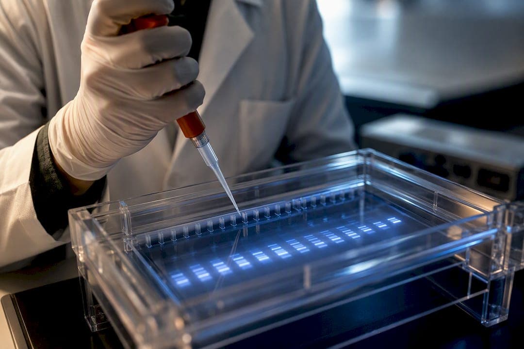
RNase H1 is the enzyme that actually does the cutting when an antisense oligonucleotide silences a gene. Once a gapmer ASO pairs with its target RNA, RNase H1 recognizes the RNA-DNA duplex and cleaves the RNA strand, provided the ASO contributes at least four consecutive DNA nucleotides to that duplex. That single structural requirement is why gapmer architecture exists at all, and why enzyme abundance and target accessibility, not just binding affinity, decide whether an ASO campaign succeeds. Design around a sufficient DNA gap and an accessible, exonic target, and cytoplasmic knockdown follows fast.
TL;DR:
- RNase H1 recognizes and cleaves RNA within a DNA-RNA duplex when at least four consecutive DNA nucleotides are present in the hybrid.
- Variability in RNase H1 abundance across cell types and its localization affects the efficiency and kinetics of ASO-mediated knockdown.
- Accurate mechanism confirmation with RISC inhibitors and RNase H1 modulation controls is essential to distinguish true RNase H1 activity from other pathways.
- In vitro assays should optimize metal ion conditions, preferably Mg2+, to reflect intracellular activity and avoid misleading cleavage patterns seen with Mn2+.
- Effective gapmer design requires a central DNA gap of 8-10 nucleotides flanked by modified wings, with accessibility and sequence context heavily influencing success.
Table of Contents
- What Is RNase H and Why Does It Matter for RNase H ASOs?
- The Catalytic Mechanism: Metal Ions and the DEDD Motif
- How Gapmer and Mixmer ASOs Recruit RNase H1
- Kinetics: Why RNase H1 Abundance Limits ASO Speed
- Sequence Preferences, Target Accessibility, and Off-Target Risk
- Running RNase H Cleavage Assays: A Practical Workflow
- Turning the Mechanism Into a Design Workflow
- What This Mechanism Doesn't Solve Yet
- How Hopeatrarelabs Applies RNase H Mechanistic Insight to Patient Programs
- What Researchers Consistently Underestimate
- How Hopeatrarelabs Can Help With Custom ASO Development
- Sources
- FAQ
What Is RNase H and Why Does It Matter for RNase H ASOs?
Human cells carry two distinct RNase H enzymes, and confusing them is one of the more common mistakes new ASO researchers make. RNase H1 is a compact, monomeric enzyme. It's the one that matters for antisense pharmacology, because it recognizes and cleaves the RNA strand within an RNA-DNA hybrid wherever the DNA-like segment runs at least four nucleotides long, cutting roughly 7 to 10 nucleotides in from the 5' end of that duplex region, according to detailed kinetic work on the RNase H1 mechanism.
RNase H2, by contrast, is a heterotrimeric complex built for a different job entirely. Its primary role is genome maintenance. It removes misincorporated ribonucleotides from DNA and resolves R-loops that form during replication and transcription. Mutations in RNase H2 subunits cause Aicardi-Goutières syndrome, a severe autoinflammatory condition, which tells you how tightly this enzyme is tied to genomic integrity rather than RNA turnover in the way RNase H1 is.
The subcellular distribution of RNase H1 explains a lot of what researchers see in knockdown experiments. RNase H1 is enriched in the nucleus, largely because of a mitochondrial and nuclear targeting sequence, but it is unambiguously active in the cytoplasm too. This dual localization is why gapmer ASOs against pre-mRNA and against mature, spliced mRNA both work, just on different timelines.
A few practical distinctions worth keeping straight when you're troubleshooting an ASO experiment:
- RNase H1 works on short RNA-DNA hybrids and needs no ATP or additional cofactor complex beyond a divalent metal ion.
- RNase H2 requires its full three-subunit assembly to function and does not substitute effectively for RNase H1 in ASO-mediated knockdown.
- RNase H1 abundance varies by cell type and cell-cycle state, which is one reason the same ASO can perform differently across cell lines.
- Other RNA turnover pathways, including RISC-mediated cleavage, can contribute at the margins, but RNase H1 remains the dominant driver of gapmer activity in essentially all published mechanistic studies.
None of this is academic trivia. If your knockdown is weaker than predicted from binding affinity alone, the first question should be whether you're actually engaging RNase H1, not whether your ASO binds tightly enough.
The Catalytic Mechanism: Metal Ions and the DEDD Motif
RNase H1's active site is built around four conserved acidic residues, commonly referred to as the DEDD motif, that coordinate divalent metal ions to position and activate a water molecule for nucleophilic attack on the RNA phosphodiester backbone. This isn't a novel biochemical trick. It's the same broad strategy used by RNase H2, retroviral RNase H domains, and several other nucleases, which is why RNase H is often treated as a model system for two-metal-ion catalysis generally, as summarized in a broad review of RNase H catalytic mechanisms.
The exact number of metal ions involved during turnover has been debated for years. The classic model calls for two metal ions, typically Mg2+ in vivo, occupying fixed sites to stabilize the transition state. More recent structural work supports a transient three-metal-ion state that appears briefly during the catalytic cycle before reverting to the two-metal resting configuration. Room-temperature X-ray and neutron crystallography studies have mapped protonation states across the active site with a level of resolution that wasn't previously possible, revealing exactly which residues gain or lose protons as the reaction proceeds, according to structural analysis of RNase H catalysis using neutron and X-ray crystallography.
Key structural finding: Neutron crystallography, unlike conventional X-ray methods, can directly visualize hydrogen and deuterium positions. That capability let researchers observe protonation changes at the DEDD residues in both the apo enzyme and the RNA-DNA hybrid-bound state, giving a much sharper picture of how the active site cycles between resting and catalytically primed conformations.
Why does any of this matter to someone running ASO experiments rather than solving crystal structures? Metal choice in your in vitro assay is not a trivial detail. Mn2+ can substitute for Mg2+ in many nuclease assays and often accelerates cleavage, but it can also relax substrate specificity and produce cleavage patterns that don't reflect what happens intracellularly, where Mg2+ predominates. A few practical consequences follow directly from the catalytic chemistry:
- Divalent cation identity and concentration change both the rate and the specificity of RNase H cleavage in cell-free assays.
- Chelators like EDTA stop the reaction cleanly by stripping the required metal ion, which is why they're the standard quench step in cleavage assays.
- Inhibitor studies need to specify which metal condition was used, because an inhibitor that blocks Mg2+-dependent cleavage may behave differently under Mn2+.
If you're interpreting an inhibitor screen or an unusual cleavage pattern, check the metal composition of your reaction buffer before you conclude the enzyme itself behaved unexpectedly.
How Gapmer and Mixmer ASOs Recruit RNase H1
The architecture that made RNase H1-dependent silencing a viable therapeutic strategy is the gapmer: a central stretch of unmodified or minimally modified DNA-like nucleotides flanked on both sides by chemically modified "wings," almost always 2'-O-methoxyethyl (2'-MOE) or locked nucleic acid (LNA) residues, all connected through a phosphorothioate (PS) backbone for nuclease resistance and improved pharmacokinetics.
The gap has to meet a specific structural threshold. RNase H1 requires a minimum of four consecutive DNA nucleotides in the duplex to recognize it as a valid substrate, a finding established through direct kinetic dissection of the cleavage mechanism. Below that threshold, the enzyme simply doesn't engage. Above it, published designs commonly use gaps in the 5 to 10 nucleotide range, with a 10-nucleotide central gap flanked by 5-nucleotide wings (the widely used "5-10-5" architecture) representing one of the most common configurations in current ASO design tools and protocols.

Mixmers complicate the tidy gapmer story in an interesting way. A mixmer interleaves modified and unmodified nucleotides throughout the sequence rather than concentrating DNA in a single central block, and you'd expect that arrangement to be a poor RNase H substrate. Experimental data say otherwise in certain contexts. One mechanistic study comparing mixmer and gapmer designs targeting the same allele-specific site found the mixmer duplex achieved roughly 65% cleavage in an in vitro RNase H assay, compared to about 14% for the gapmer duplex under the same conditions, according to mechanistic work on allele-specific mixmer and gapmer ASOs. That same study also found that a RISC inhibitor partially restored target expression in gapmer-treated cells, pointing to a minor RNA interference contribution layered on top of the dominant RNase H1 mechanism.
That mixed-mechanism finding matters more than it might first appear. If your mechanism-of-action data doesn't cleanly separate RNase H1-dependent cleavage from RISC involvement, your specificity conclusions could be off. A gapmer producing an unexpected knockdown pattern isn't necessarily behaving unpredictably through RNase H1. It might be getting partial help from a pathway you didn't design for.
Practical takeaways for architecture selection:
- Confirm your gap length meets or exceeds the four-nucleotide minimum before troubleshooting anything else.
- Treat mixmers as a viable alternative when allele discrimination matters more than maximal knockdown magnitude, since their partial-complementarity tolerance can favor selectivity.
- Run a RISC inhibitor control (such as aurintricarboxylic acid) alongside your standard RNase H assay whenever you need a clean mechanism-of-action claim.
- Expect cleavage to occur within a fairly narrow window inside the duplex region rather than uniformly across the ASO footprint, and design your detection assay (qPCR primers, Northern probe placement) around that expectation.
Kinetics: Why RNase H1 Abundance Limits ASO Speed
Kinetic and subcellular fractionation studies give a clear picture of how fast RNase H1-mediated knockdown actually happens, and the answer depends heavily on where in the cell your target RNA lives. Exon-targeting ASOs directed at mature, spliced mRNA in the cytoplasm can reduce target transcript levels detectably within 30 minutes of transfection in some systems, based on fractionation experiments tracking RNase H1 activity in both nuclear and cytoplasmic compartments. Intron-targeting ASOs act on pre-mRNA in the nucleus, and their apparent knockdown kinetics track the natural turnover rate of that pre-mRNA species rather than the intrinsic speed of RNase H1 cleavage itself.
Statistic worth remembering: Overexpressing RNase H1 increased ASO-induced degradation rates by roughly 1.6 to 2-fold in kinetic experiments, while knocking down RNase H1 dropped degradation to near background levels seen without any ASO at all, according to the same mechanistic kinetics study. That range tells you enzyme abundance isn't a minor variable. It's often the rate-limiting step in the entire pathway.
This has real consequences for how you plan experiments and interpret dose-response curves:
- Don't assume a linear dose response across your ASO concentration range. If RNase H1 is saturating at moderate ASO concentrations, higher doses will show diminishing returns that have nothing to do with target binding.
- Sample your knockdown timecourse early and often, especially in the first few hours, if you're comparing exon versus intron targeting strategies.
- Consider RNase H1 modulation (siRNA knockdown or transient overexpression) as a standard orthogonal control when you need to prove your knockdown is genuinely RNase H1-dependent rather than an artifact of another degradation pathway.
- Cell type matters. A knockdown protocol optimized in one cell line may underperform in another simply because baseline RNase H1 levels differ.
Binding affinity gets most of the attention in ASO design discussions, but affinity alone doesn't predict knockdown speed. Two ASOs with nearly identical binding thermodynamics can produce very different kinetic profiles if one recruits RNase H1 more efficiently or targets a site with better accessibility. That distinction is exactly why sequence and structural determinants deserve their own close look.
Sequence Preferences, Target Accessibility, and Off-Target Risk
RNase H1 does not treat every DNA-RNA duplex equally, even when all of them clear the four-nucleotide minimum. Sequence preference profiling using assays like H-SPA (RNase H sequence preference assay) has shown that local sequence context around the cleavage site materially affects cutting efficiency, independent of overall thermodynamic binding strength, according to work on RNase H sequence preferences and antisense efficiency. Designing purely against a binding-energy calculator and ignoring cleavage-site sequence context is a common way to end up with an ASO that binds well but underperforms functionally.
RNA secondary structure is the other major variable, and it's arguably the more consequential one in practice. A perfectly designed gapmer against a structurally occluded region of an RNA can simply fail to hybridize efficiently, regardless of how well the sequence itself would perform against an open target. Computational structure prediction tools combined with experimental accessibility probing, such as RNase H mapping or SHAPE-based structure probing, remain the most reliable way to identify genuinely accessible windows rather than relying on sequence complementarity scores alone. Hopeatrarelabs' own comparison of antisense oligonucleotide design approaches walks through how accessibility screening feeds into gapmer site selection.
Off-target effects in RNase H-dependent ASOs come from two distinct sources, and treating them as the same problem leads to the wrong fix:
- Partial complementarity off-targets, where the ASO tolerates enough mismatches against an unintended transcript to still form a cleavable duplex, typically require BLAST-style transcriptome screening against the full sequence, not just the perfect-match target.
- Immunostimulatory motifs, particularly unmethylated CpG dinucleotides within the PS backbone context, can trigger innate immune activation independent of any RNase H mechanism at all, and need to be flagged and removed during sequence design rather than caught after the fact in a toxicity screen.
Pro Tip: Run your candidate gapmer sequences through an off-target and liability screen (G-quadruplex motifs, CpG content, self-complementarity) before you order synthesis, not after your first cell-based assay comes back messy. It's far cheaper to filter in silico than to troubleshoot a confounded result. Hopeatrarelabs' off-target assessment workflow for ASO labs outlines an SPR-based confirmation step that catches binding artifacts sequence screening alone can miss.
Running RNase H Cleavage Assays: A Practical Workflow
Validating that your ASO actually works through RNase H1, rather than some other silencing pathway, comes down to a fairly standardized in vitro workflow that most published mechanistic studies follow with minor variations.
- Anneal the duplex. Combine your ASO with a complementary RNA target (synthetic oligo or in vitro transcribed RNA) in an appropriate buffer, heat to denature, and cool slowly to allow specific hybridization.
- Incubate with RNase H. Add purified RNase H enzyme, commonly a commercial preparation such as NEB's RNase H (catalog M0297), which has shown up repeatedly in mechanistic ASO literature as a standard reagent for in vitro cleavage assays, including the allele-specific mixmer and gapmer study cited earlier. Incubate at 37°C for a defined time course, typically sampling at several time points from a few minutes out to an hour.
- Stop the reaction with EDTA. Chelating the divalent metal cofactor halts catalysis immediately, giving you clean time points rather than a continuously progressing reaction.
- Resolve on a denaturing gel. Run samples on a TBE-based polyacrylamide or agarose gel depending on fragment size, then visualize cleavage products against uncleaved substrate.
For cell-based confirmation, transfect your ASO and sample across a timecourse rather than a single endpoint. Because exon-targeting ASOs can show detectable knockdown within 30 minutes in cytoplasmic fractions, while intron-targeting effects lag behind pre-mRNA turnover, a single 24 or 48-hour endpoint will often obscure the actual kinetic profile. Subcellular fractionation into nuclear and cytoplasmic pools before RNA extraction lets you attribute knockdown to the correct compartment rather than assuming a uniform effect.
When interpreting gel results, look for cleavage products of the expected size given your predicted RNase H1 cut site, roughly 7 to 10 nucleotides in from the duplex's 5' end. A diffuse smear rather than discrete bands often indicates degradation from a nonspecific nuclease contaminant rather than genuine RNase H1 activity, and a RISC inhibitor or RNase H1 knockdown control is the cleanest way to confirm which pathway you're actually measuring.
Turning the Mechanism Into a Design Workflow
Everything above collapses into a fairly compact set of design rules once you're actually building a gapmer for a specific target.
Start with architecture. A central DNA gap of at least four nucleotides is the non-negotiable floor, but published and commercially deployed designs generally use 8 to 10 nucleotides in the gap for reliable RNase H1 engagement, flanked by wings of 2'-MOE or LNA chemistry that boost binding affinity and nuclease resistance without themselves being cleavable substrates. A phosphorothioate backbone throughout remains standard for pharmacokinetic stability in vivo, though it does carry known protein-binding liabilities worth screening for separately.
Target selection comes next, and the exon-versus-intron decision should be deliberate rather than incidental:
- Target exonic, mature mRNA sequence when you need the fastest possible cytoplasmic knockdown for a functional screen or an acute experimental timeline.
- Target intronic or pre-mRNA sequence when you're aiming to modulate splicing outcomes or when nuclear-localized transcripts are your actual biological target.
- Favor sites that appear in multiple transcript regions or isoforms if your goal is knockdown breadth across splice variants, but verify this doesn't inadvertently reduce specificity against related paralogs.
- Confirm accessibility experimentally wherever possible rather than trusting structure prediction alone, since predicted and actual accessibility can diverge meaningfully at individual sites.
Pro Tip: Build your optimization pipeline as a funnel, not a single-pass filter: in silico screening (thermodynamics, off-target BLAST, liability motifs) to generate a shortlist, then a small-scale in vitro RNase H assay to confirm cleavage, then cellular kinetics with fractionation, then a transcriptome-wide off-target check on your top one or two candidates. Running the expensive transcriptome-wide assay on ten candidates instead of two is the single most common waste of time and budget in early ASO campaigns.
The full sequence, roughly, looks like this: computational filtering for accessibility, thermodynamics, and liability motifs; a cell-free RNase H cleavage assay to confirm the candidate is a genuine substrate; cellular transfection with timecourse sampling and nuclear/cytoplasmic fractionation; RNase H1 modulation controls (knockdown and overexpression) to confirm mechanism; and finally a transcriptome-wide off-target assessment on your leading candidates before committing to further development. Hopeatrarelabs' overview of ASO development essentials for genetic disease therapies covers how these steps connect to translational planning for programs aimed at eventual clinical use.
What This Mechanism Doesn't Solve Yet
RNase H1-dependent gapmer chemistry is mature science, but it isn't a finished picture, and pretending otherwise sets up unrealistic expectations for new researchers entering the field.
Delivery remains the dominant bottleneck for translating cell culture success into in vivo efficacy, particularly for tissues outside the liver and central nervous system where unconjugated or lightly conjugated ASOs distribute well. Pharmacokinetic and pharmacodynamic disconnects, where plasma exposure looks reasonable but tissue-level target engagement doesn't follow proportionally, are common enough that PK/PD modeling has become a standard part of preclinical ASO programs rather than an afterthought.
A few other open issues are worth flagging honestly:
- Enzyme saturation at higher ASO doses produces non-linear dose-response curves that complicate simple potency comparisons across candidate sequences.
- Allele-specific targeting, where a single nucleotide difference needs to drive selective cleavage, remains genuinely difficult, and the mixmer strategies discussed earlier are a partial solution rather than a complete one.
- Comprehensive in vivo sequence-preference maps for RNase H1 across diverse tissue and cell types don't yet exist at the resolution researchers would like.
- How individual variation in human RNase H1 expression or activity affects patient-to-patient ASO response is still an underexplored question with direct relevance to personalized dosing strategies.
- The dynamics of metal cofactor availability inside living cells, as opposed to controlled in vitro buffer conditions, remain poorly characterized despite the detailed structural picture available for the isolated enzyme.
None of these gaps invalidate the core mechanism. They just mean the field still has real work to do translating a well-understood biochemical reaction into consistently predictable therapeutic outcomes.
How Hopeatrarelabs Applies RNase H Mechanistic Insight to Patient Programs
The mechanistic details covered here aren't abstract for Hopeatrarelabs. They shape how custom ASO candidates get tested against a specific patient's own biology. Working from patient-derived induced pluripotent stem cell (iPSC) models, often paired with CRISPR-edited isogenic controls to isolate the effect of a specific mutation, Hopeatrarelabs runs custom ASO candidates through cleavage validation and cellular kinetics as part of a broader parallel screen that also evaluates thousands of FDA-approved compounds and gene therapy feasibility side by side.
The core translational question is never just "does this ASO bind the target?" It's whether RNase H1 actually engages the duplex efficiently in that patient's own cells, at a rate fast enough to matter clinically, without triggering off-target liabilities in a genetic background that may itself be unusual.
That's the practical value of building disease models from a patient's actual cells rather than a generic cell line: RNase H1 abundance, target accessibility, and splicing patterns can all vary with genetic background, and a patient-specific model captures that variability directly instead of assuming it away. Programs contracted through Hopeatrarelabs typically move through disease modeling, parallel treatment screening, and results translation, with the goal of giving families, physicians, and biopharma partners a scientifically grounded answer rather than a speculative one. More detail on how this applies specifically to custom ASO work is available in Hopeatrarelabs' overview of custom ASOs in rare disease treatment.
What Researchers Consistently Underestimate
The biggest mistake I see in ASO campaigns isn't a design flaw in the gapmer itself. It's sequencing the experiments in the wrong order. Teams often spend weeks optimizing binding affinity and backbone chemistry before ever confirming that RNase H1 is engaging the target efficiently in their specific cell type, then get confused when a thermodynamically "perfect" ASO underperforms.
Measure target accessibility and run a basic RNase H1-limited kinetics check early, before you've committed to a final architecture. It's cheap, it's fast, and it tells you whether you're solving a binding problem or an enzyme-engagement problem, which are fixed with completely different design changes.
Orthogonal mechanism controls aren't optional if you're making a mechanism-of-action claim in a publication or a regulatory filing. RNase H1 knockdown and overexpression, alongside a RISC inhibitor control, cost little relative to the credibility they add. The mixmer data discussed earlier make clear that assuming pure RNase H1 dependence without checking is a real risk, not a theoretical one.
Know when the work has outgrown your bench. Patient-specific modeling, high-throughput parallel screening across drug libraries and gene therapy options, and transcriptome-wide off-target profiling are resource-intensive enough that partnering with a specialized lab often beats trying to scale internally, particularly for ultra-rare disease programs with a single patient's cells to work from.
— John
How Hopeatrarelabs Can Help With Custom ASO Development
If the design principles above raised more questions than you can resolve with a standard academic lab setup, that's precisely the gap Hopeatrarelabs was built to close. Hopeatrarelabs runs custom ASO development alongside patient-specific iPSC disease modeling and parallel screening against thousands of FDA-approved drugs and gene therapy options, all built from a patient's own cells rather than a generic proxy cell line.

That patient-specific foundation matters most for ultra-rare and undiagnosed genetic diseases, where no approved treatment exists and generic cell lines simply can't capture the relevant genetic background. Families, physicians, foundations, and biopharma partners typically start by scoping the specific disease and gene target, arranging a sample collection plan for iPSC derivation, and setting a realistic timeline for parallel drug, ASO, and gene therapy screening given the biology involved. If you're facing a diagnosis where standard treatment options have run out, reach out to Hopeatrarelabs to discuss whether a personalized screening program fits your situation, and see how the process moves from initial scoping through to translated results.
Sources
- The rates of the major steps in the molecular mechanism of RNase H1-dependent antisense oligonucleotide induced degradation of RNA
- Structural insights into RNase H catalytic mechanism from room-temperature X-ray and neutron crystallography of apo- and RNA/DNA hybrid-bound enzyme
- The catalytic mechanism, metal dependence, substrate specificity, and biodiversity of ribonuclease H
FAQ
What Is RNase H Used For?
RNase H degrades the RNA strand within an RNA-DNA hybrid, a function essential to DNA replication (removing RNA primers), genome maintenance, and, in antisense pharmacology, cleaving target mRNA or pre-mRNA when a gapmer ASO forms a duplex with it.
Do Humans Have RNase H?
Yes. Human cells express two forms, RNase H1 and RNase H2, and RNase H1 is the enzyme responsible for cleaving RNA in gapmer ASO-directed knockdown, as established in detailed kinetic studies of the mechanism.
What Diseases Can ASOs Treat?
RNase H-dependent ASOs are approved or in development for conditions including spinal muscular atrophy, certain hereditary transthyretin amyloidosis cases, familial hypercholesterolemia, and a growing list of ultra-rare and undiagnosed genetic diseases where a custom-designed ASO can target a patient-specific mutation.
How Much Does Antisense Oligonucleotide Therapy Cost?
Published ASO drug pricing varies enormously by indication, dosing schedule, and whether the therapy is commercially approved or an individualized/investigational program, so no single figure applies across the field; individualized programs are typically scoped and quoted based on the specific disease and development pathway involved.
What's the Minimum DNA Gap a Gapmer Needs for RNase H1 Activity?
RNase H1 requires at least four consecutive DNA nucleotides in the duplex to recognize it as a cleavable substrate, though most practical gapmer designs use gaps of 8 to 10 nucleotides for reliable activity.